


 , Bashuli Acharyya and Ethiraj Selvarajan
 The surfaces of medical devices can quietly become breeding grounds for resilient bacterial colonies, which pose a significant risk to patients. These microorganisms form complex communities embedded within a self-produced extracellular matrix known as the extracellular polymeric substance, which confers enhanced resistance to antimicrobial agents and the host’s immune responses. Thus, developing methods to degrade the EPS is crucial for improving infection control in medical implants. Studies reveal that enzymes can inhibit biofilm formation by targeting essential components of the EPS, including exopolysaccharides, extracellular DNA, and proteins. Enzyme immobilization enhances enzyme reusability and stability, thereby improving biofilm eradication capabilities. They exhibit superior catalytic activity, turnover rates, and specificity in comparison to free enzymes. This systematic review examines literature from the past fifteen years (2010-2024), focusing on articles detailing enzyme immobilization techniques and their application against biofilms in the context of biomaterials and tissue engineering scaffolds. The enzyme-polysaccharide complex appears especially well suited to promote biofilm dispersal. Integrating enzyme immobilization with tissue engineering offers the possibility of improved development of infection-resistant tissue constructs.
Biofilm Eradication, Enzyme Immobilization, Antibiofilm Enzymes, Tissue Engineering, Infection-resistant Implants
In the ever-evolving field of healthcare, medical devices have revolutionized treatment, yet they often face an unseen adversary: bacterial biofilms. Biofilms are intricate networks of microorganisms that are highly resistant to antibiotics due to their robust extracellular matrix. In the field of tissue engineering, biofilm growth on any biomaterial significantly hinders the process of cell attachment, compromises the efficiency of the scaffolding material, or contributes to ongoing infection. Conventionally, antibiotic treatments are inept because EPS presents a shielding ability. Therefore, newer approaches toward developing strategies are being introduced against biofilm-mediated challenges with the tissue constructs.1
One useful approach to break down biofilms is the use of specialized enzymes capable of dismantling the basic polysaccharides, proteins, and extracellular DNA that can have built up to form the biofilm. This way, by targeting these key components, the enzymes can cause a shift in biofilm structure that can make it more susceptible to treatments. Some polysaccharide-degrading enzymes have also been reported to be effective at dispersing biofilms through structural matrix disruption, within which microbial cells are embedded. For example, glycoside hydrolases degrade exopolysaccharides, resulting in the dispersal of the biofilm with increased sensitivity towards antimicrobial agents.1
Polysaccharides, both in their natural and modified forms, serve as effective matrices for enzyme immobilization owing to their inherent biocompatibility, biodegradability, and tunable functionalities. This approach enhances enzyme stability and catalytic activity while enabling controlled enzyme release, rendering it particularly advantageous for biomedical applications. For example, immobilization on the biodegradable, natural polysaccharide chitosan has permitted biofilm degradation through enzyme-impregnated biopolymers to effectively accelerate healing in wounds.2
More recent breakthroughs in immobilization technologies have further expanded the inventory of available methodologies and utilization and led to the building of stronger conjugates of enzymes and polysaccharides for degrading biofilm.3
In summary, enzyme-immobilized polysaccharides play an important role as a multifaceted approach in the challenge related to biofilm in tissue engineering. Given the properties of the polysaccharide carriers and the action mechanisms exerted by biofilm-degrading enzymes, one can achieve more durable and clinically effective tissue constructs.
Biofilms in biomedical applications
Structure and Composition
Biofilms are microorganism communities that adhere to a surface and develop a self-sustaining polymeric matrix known as the extracellular polymeric substance (EPS). The EPS acts as some kind of shell to preserve the microbes and allow their mutual coexistence, forming an ecosystem within the biofilm (Figure 1). It is crucial in biofilm formation for providing the cohesion and adhesion forces needed to maintain its structure. It also serves as a scaffold for cell-to-cell communication.4
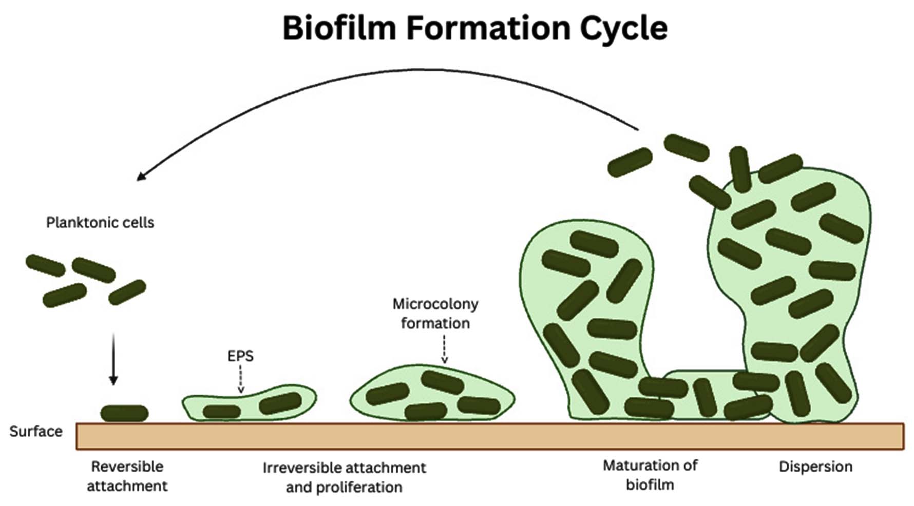
Figure 1. Overview of biofilm development from planktonic cell attachment to mature EPS-embedded biofilm (Source: https://BioRender.com)
Furthermore, it functions as a defensive barrier against threats such as oxidizing biocides, antibiotics, UV radiation, and the host immune system.4
The EPS matrix is a composition of exopolysaccharides, proteins, and nucleic acids. A considerable portion of the EPS is more or less strongly hydrated, apart from cellulose, which is hydrophobic.5 This matrix surrounds the cells and allows them to communicate with one another through gene exchange and metabolic signals.6 Under some circumstances, biofilms may become fossilized (stromatolites) due to the strength of this matrix.
Biofilm-associated challenges
Biofilms were initially recognized as biofouling agents about a century ago. However, the identification of large aggregates of Pseudomonas aeruginosa cells in sputum and lung tissue from patients with chronic cystic fibrosis in the early 1970s was instrumental in establishing the concept of biofilm infections and their significance in medical research.7 Biofilms as agents damaging medical instruments like polymer-based cardiac pacemakers and catheters.8,9 These findings were noted as modern challenges in medicine brought further support toward enhanced strategies to prevent and manage biofilm-related complications.10 The term “polymer-associated infection” emerged to describe biofilm-related infections, a consequence of the widespread use of medical devices in healthcare environments.
The primary microorganisms implicated in biofilm formation on indwelling medical devices include “Gram-positive bacteria such as Enterococcus faecalis, Staphylococcus aureus, Staphylococcus epidermidis, and Streptococcus viridans, alongside Gram-negative bacteria like Escherichia coli, Klebsiella pneumoniae, Proteus mirabilis, and Pseudomonas aeruginosa, and yeasts”.11 Infected implants are believed to operate as a reservoir for infection of the surrounding tissue due to the intracellular survival of bacteria.12 As a result, infections linked to biomedical devices are difficult to manage with antibiotic treatments and are resistant to the body’s immune defenses.13
Biofilm degradation
The formation of biofilms can be prevented and bacterial sensitivity increased by anti-biofilm tactics that target specific elements of bacterial cells and biofilm structure. Competitive adhesin inhibitors prevent bacterial attachment and biofilm growth by targeting pili and fimbriae on the cell membrane. Quorum-sensing inhibitors (QSIs) and acylase enzymes affect communication routes by interrupting ligands such as acyl-homoserine lactones. These molecules play an important role in biofilm creation. Additionally, iron chelators decrease the iron supply, which is key for biofilm formation and steadiness2 (Figure 2). Enzymes have emerged as potent and effective agents due to their ability to degrade biofilms and liberate microorganisms into a more susceptible planktonic state.14 Enzymatic therapies as more advantageous than other biofilm dispersal techniques.15 Dispersal agents are therefore used to increase the bacteria’s access to antimicrobials and host immune cells, thereby improving therapeutic outcomes.16 High specificity and efficacy against the targeted biofilms are obtained at comparatively low concentrations with the use of biofilm-dispersing enzymes, which act more significantly against both developing and existing biofilms.17,18
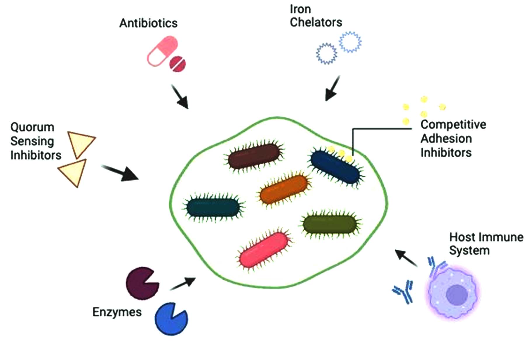
Figure 2. Schematic illustration of the major strategies employed for biofilm dispersion and bacterial eradication (Source: https://BioRender.com)
Polysaccharides as an immobilization platform
Polysaccharides serve well as medicinal agents and enzyme carriers due to several factors,19,20 such as (a) their abundance in the environment, and well-established techniques for extracting them from biotic sources are readily available.21 (b) Despite their eco-friendly nature, polysaccharide-based biopolymers remain underutilized.22 (c) These polysaccharides can be modified using both synthetic approaches and protein-related techniques to maximize their functionality.23 Glycans offer a diverse range of properties that can improve many types of carrier systems. For instance, ionic glycans possess increased sensitivity and pH, which leads to improved catalytic reaction in the enzyme-carrier procedure.24 Because of these characteristics, they are an ideal biomaterial for developing controlled enzyme delivery systems. This system is designed to release the encapsulated enzyme at a target location and specific time during a pharmacological process, in response to a specific biological catalyst.24
Carriers of immobilized enzymes can be divided into two types: organic and inorganic
In the instance of organic carriers, only a few synthetic polymers and some natural polysaccharides, such as chitin, chitosan, pectin, cellulose, and alginate, are used.25 Polysaccharides as enzyme carriers have gained much attention from researchers globally due to their numerous advantages, like facile chemical modifiability post-immobilization and easy control of the enantioselectivity, chemoselectivity, and regioselectivity characteristics of enzymes for a particular application after the enzyme immobilization process. It aids in enhancing the yield of the end product, reversing an undesirable reaction, and controlling the homogeneity of a reaction.25
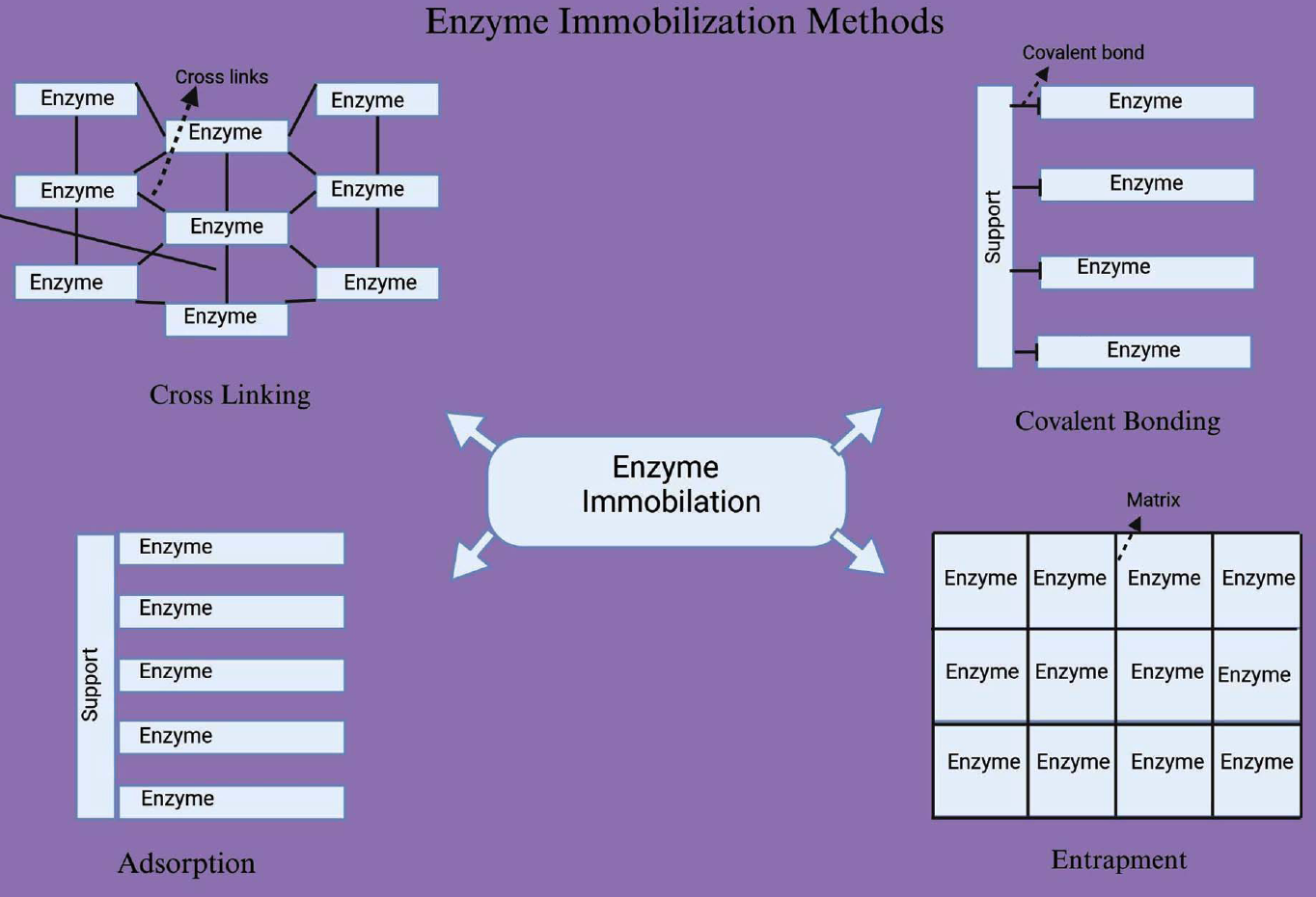
Figure 3. Different enzyme immobilization methods showing physical (adsorption, entrapment) and chemical (covalent bonding, cross-linking) approaches for improved enzyme stability and reusability
Mechanism of enzyme immobilization
Biochemical and biophysical research has aimed to immobilize enzymes to increase their stability and activity throughout the past few decades.26 Immobilized enzyme catalysts have significantly enhanced industrial processes’ technical performance, leading to higher economic efficiency and productivity.27 Enzyme immobilization strategies are broadly categorized into two types: physical and chemical28 (Figure 3). Physical methods rely on fragile interactions such as affinity binding, ionic binding, van der Waals forces, hydrophobic interactions, hydrogen bonds, and entrapment of the enzyme within the support.29,30 On the other hand, chemical techniques are characterized by the formation of covalent bonds between the enzyme and the support material through ether, thioether, amide, and carbamate linkages.29 The four main techniques for immobilizing enzymes are categorized as follows:
- Adsorption; 2. Entrapment; 3. Covalent Bonding and 4. Cross-linking
Adsorption
Among the simplest forms of immobilization is the adsorption of enzymes on the solid surfaces of biosensor transducers. Adsorption mechanisms are based on weak bonds such as van der Waals forces, hydrogen bonds, and hydrophobic interactions.27 Under ideal situations that allow for enzyme activity, the solid support is in proximity for a determined amount of time. Some popular supports for enzyme immobilization through adsorption include collagen, chitin, cellulose, and polyethylene glycol (PEG).25 After that, the surface is cleaned with a buffer to get rid of the unadsorbed enzyme molecules. Since there is no functionalization of the support involved, immobilization by adsorption is a straightforward, cost-effective, reagent-free technique that is often non-destructive to enzyme performance.26 The disadvantages of this method include the possibility of enzyme desorption or leaching due to temperature difference, pH, or ionic strength because the enzymes are only weakly physically bonded to the support. In addition, nonspecific adsorption of other proteins or chemicals on the transducers’ surface can contaminate and block signals, making adsorbed enzyme biosensors unstable both during operation and storage.31
Entrapment
Entrapment for enzyme immobilization, involves enclosing the enzymes in a matrix; this could be a material’s lattice structure, or polymer membranes.32-34 It holds the enzyme together, but allows the substrate and products to pass through.35 Because the enzyme does not chemically react with the polymer, the enzyme is protected by the polymer, limiting denaturation.36,37 Because this appeals to an immobilization method, it is possible to modify the encasing material, creating the ideal environment for the enzyme. The ideal environment could contain ideal pH, polarity, and amphiphilicity. These characteristics can be adjusted with a variety of polymers, sol-gels, polymer /sol-gel /inorganic hybrid composites.38-41 Research identifies as acceptable polpes: alginate, carrageenan, collagen, polyacrylamide, gelatin, silicone rubber, polyurethane and poly(vinyl) alcohol with styrylpyridinium substituent.42 Alginates are one of the most commonly used polymers because of their “relatively low toxicity and mild gelling properties”.36 Even with the benefits of entrapment discussed above, this technique has a limited usefulness because of the mass transfer limitations of the substrate/analyte to the enzyme active site.43 Additional drawbacks include deactivation during immobilization, potential enzyme leaking, diffusion barriers, and wearing down of the support matrix.28
Covalent Bonding
Covalent bonds is one of the most popular approaches for immobilizing enzymes because they make strong, stable complexes between the enzyme and support matrix. The enzyme coupling is usually accomplished by functional groups, such as the side chains of lysine (ε-amino group), cysteine (thiol group), and aspartic and glutamic acids (carboxylic group),44 imidazole, a lot of phenolic groups, etc.45 The process of enzyme coupling to a stiff matrix usually involves two steps: surface activation using appropriate linkers (such glutaraldehyde or carbodiimide) and covalent enzyme binding to the activated support.42 The surfaces are bi-functional (glutaraldehyde/carbodiimide) pre-linking molecules, in which part of the prewashed and condensed support make covalent bonds to the exterior of the enzyme(s), while the other half become covalently linked to the enzyme through the functional groups. The first group, used for the creation of self-assembled monolayers (SAMs), became highly ordered by binding to the surface. The second group linked to the pre-activated support tightly enough to form a covalent bond with the enzyme. The size, shape, and composition of the carrier material as well as the type of coupling mechanism affect the covalently coupled enzyme’s activity.45
Cross-linking
Cross-linking is an irreversible enzyme immobilization technique that relies on cross-linking reagents to form covalent bonds between enzyme molecules.32,46,47 The multipoint binding reagents link enzyme molecules into three-dimensional crosslinked aggregates.48,49 The reaction mixture contains an immobilized enzyme that has not been bound. Cross-Linking Enzyme Aggregate (CLEA) and Cross-Linking Enzyme Crystal (CLEC) are two common approaches to cross-linking immobilization.42 Glutaraldehyde, a common cross-linking agent, is employed in both methods to establish covalent bonds between the free amino groups present on the lysine residues of adjacent enzyme molecules.43,48,49 In the CLEC method, glutaraldehyde is applied after the enzyme has been crystallized. Enzymes immobilized using CLECs are generally more stable and effective than their untreated forms, as they often demonstrate mechanical properties. Glutaraldehyde usage, however, may cause significant enzyme alterations, including loss of function and potential structural abnormalities.70 Therefore, to minimize the significant alteration of enzymes as they are immobilized, inert proteins such as gelatin and bovine albumin need to be incorporated into the process.71
Enzyme polysaccharide systems for biofilm degradation
Extracellular enzymes have the ability to effectively disrupt bacterial biofilms by targetting the main structural components of the extracellular polymeric substance (EPS) specifically: exopolysaccharides, extracellular DNA, and extracellular proteins.18 The three enzyme classes or categories promote bacterial cell transition from a surface-adhering state to a planktonic state by affecting the stability of the biofilm’s polysaccharide matrix. This transformation makes the bacteria more vulnerable to antibiotics and the host’s immune system.72 Therefore, to harness this property, biofilm-dispersing enzymes can be produced in large quantities through laboratory approaches like recombinant overexpression in vectors and then applied externally to microbial populations for efficient biofilm degradation.18
Biofilm disruption is done using three main groups of enzymes: proteases,73 deoxyribonucleases74,75 and glycoside hydrolases.76 The mechanism and application of these biofilm-dispersing enzymes are reviewed in Table.
Table. Enzymes Targeting Biofilm Components: Mechanisms and Applications
No. |
Enzyme |
Type |
Biofilm Target |
Mechanism |
Key Experimental Findings |
Ref. |
|---|---|---|---|---|---|---|
1 |
Proteinase K |
Serine Protease |
Exoproteins in biofilms |
Breaks peptide bonds near carboxylic groups of aromatic/aliphatic amino acids |
In vitro: Dispersed 24-h and 48-h old S. aureus biofilms. Co-treatment: Synergistic degradation of preformed Listeria monocytogenes biofilms when combined with DNase I. |
50-52 |
2 |
Trypsin |
Serine protease |
Biofilms on teeth and wounds |
Cleaves peptide bonds on the carboxyl side of lysine and arginine |
In vitro: Alone, reduced biomass of 24-h old P. aeruginosa and E. faecalis biofilms. Synergy: Co-administered with Pepsin and Carvacrol, achieved complete dispersal of mature P. aeruginosa and E. faecalis biofilms on abiotic surfaces. |
53-55 |
3 |
Pepsin |
Endopeptidase |
Biofilms on polystyrene surfaces |
Cleaves Phe and Leu residues; inhibited by His, Lys, Arg, Pro. |
In vitro: Reduced biomass of 24-h old P. aeruginosa and E. faecalis biofilms. Synergy: Must be co-administered with Trypsin and Carvacrol for full eradication. |
55-57 |
4 |
DNase I |
Nuclease |
eDNA in biofilms |
Breaks down eDNA reducing biofilm structure and bacterial viability |
In vitro: Disrupted formation of both mono- and poly-microbial biofilms, reducing biofilm biomass and increasing antibiotic susceptibility. In vivo: Recombinant human DNase I (rhDNase) is clinically applied in cystic fibrosis patients to reduce sputum viscosity. |
58-60 |
5 |
Nucleases Xds & Dns |
Nucleases |
DNA in biofilms |
Degrade circular and linear DNA; accelerate biofilm formation when absent |
Clinical Relevance: Deletion of encoding genes in Vibrio cholerae results in increased biofilm formation, indicating a strong dispersal function. |
61,62 |
6 |
NucB |
Endonuclease |
eDNA in biofilms |
Non-specific cleavage of eDNA |
Clinical Relevance: Degraded preformed biofilms of staphylococci and streptococci isolated from chronic rhinosinusitis infections. |
63,64 |
7 |
Dispersin B |
Glycoside hydrolase (GH20) |
dPNAG in biofilm matrices |
Hydrolyzes dPNAG using a substrate-assisted mechanism |
In vitro: Effective against S. aureus, A. baumannii, and K. pneumoniae biofilms. In vivo/ Clinical Relevance: Has been commercialized as a wound gel for accelerated healing of infected and non-infected dermal wounds. |
65-67 |
8 |
Alginate lyase |
Glycoside hydrolase |
Alginate in biofilms |
Breaks down alginate, particularly polyM/G activity enzymes |
In vitro: Enzymes with polyM/G activity are effective in destroying preformed mature biofilms. Synergy: Marine alginate lyase (AlyP1400) enhanced the bactericidal activity of Tobramycin by modulating efflux antibiotic resistance. |
68,69 |
Application in tissue constructs
Biofilm-dissolving, enzyme-functionalized, and polymer-based scaffolds are transforming tissue engineering by promoting tissue incorporation and inhibiting microbial colonization. As biofilm formation can disrupt healing, the vital role of biofilm-reactive scaffolds in skin, vascular, and bone tissue regeneration.18 This is consistent research emphasize the use of enzyme-containing polysaccharide-based composites to remove biofilms from chronic wounds and create a sterile environment for the growth of new tissue.3,77 The effectiveness of enzyme-functionalized scaffolds, adopt a more nuanced stance, contending that localized antibacterial effects via polysaccharide-enzyme drug delivery systems may lessen systemic side effects and improve therapeutic outcomes.78 This presents a shift from merely biofilm clearance to a controlled antibacterial response that minimizes potential adverse effects.
In the context of bone and cartilage regeneration, biofilm-resistant constructs play a major role in maintaining an aseptic environment crucial for grafting success.79 Their claim underscores the necessity of biofilm-resistant scaffolds, that vascular graft infections, while mitigated through infection prevention programs and endothelialization support, also depend on enzyme-immobilized polysaccharides for success.80 This indicates that while biofilm resistance is essential, additional factors beyond scaffold functionality play a role in vascular graft integration. The subject of dental implants has become increasingly debatable. Enzyme-coated polysaccharide scaffolds assist in prolonging the lifespan of implants by preventing bacterial adhesion and proliferation.1 While this is in line with previous claims of biofilm-resistant benefits, it raises the question of whether other antibacterial strategies, without enzyme-based coatings, could yield similar or superior outcomes.
Overall, while enzyme-functionalized, biofilm-resistant scaffolds are a cutting-edge new product in regenerative medicine, how effective they are varies with the application environment. Although most studies are in favor of their ability to enhance healing and improve implant survival rates, other studies introduce complications, raising questions as to whether biofilm resistance is always optimal.
Challenges and limitations
Systems for biofilm degradation that are based on enzymes face numerous challenges that must be dealt with before they can be successfully applied in biomedical fields. One of the main issues is the stability and activity of the enzymes since, during physiological processes, they are often deactivated due to denaturation or interactions with host factors.81 As evidenced by recent studies where nanostructured carriers preserved enzyme activity under physiological stress, future research may concentrate on encapsulating enzymes in nano-polysaccharide hybrid matrices to improve thermal and conformational stability.71
The next big hurdle is associated with scalability and cost because the production of polysaccharide-enzyme systems, on a large scale, is still very expensive and resource-heavy. Even developing cheaper synthesis and extraction technologies for sustainable polysaccharide polymers could serve as a remedy for this challenge.82 Cost-effective cross-linked enzyme aggregates (CLEAs) and continuous-flow bioreactor designs have demonstrated promise for industrial-scale enzyme immobilization, which could reduce production costs and increase reusability.83
Also, the heterogeneous biological nature of the biofilm renders the design of a universal enzyme system incompetent. It is crucial to customize the enzyme formulations relative to targeted specific biofilm compositions along with the developmental stages of biofilms for the overall functioning of enzymes.84 According to recent modeling studies, degradation efficiency could be greatly increased by customizing immobilized enzyme systems to particular EPS compositions using directed evolution and computational enzyme design.85
The inherent risk of potential immunogenicity remains a concern, especially for sensitive patients, since both enzymes and polysaccharides trigger the immune response. To attenuate these effects, biocompatible and hypoallergenic materials be used.86 Equally contributing to the challenge is the fact that regulatory challenges encompass the clinical applicability of enzyme-polysaccharide systems that need stringent approval requirements. By standardizing test procedures, the market authorization process shall be streamlined.87 Other obstacles include the short half-life of enzymes, which warrants schemes like repetitive reapplication or formation of reservoir-based enzyme delivery systems.88 Finally, there are still some administrative and operational challenges that result from the combination of enzyme-polysaccharide systems with advanced technologies such as 3D bioprinting, nanotechnology, and smart materials. There is an exigent need for novel approaches to bridge such gaps and enhance the functional potentials of the enzyme-immobilized constructs.71 The integration of enzyme-functionalized bioinks into 3D-bioprinted scaffolds can facilitate spatial control of enzyme activity, bridging the gap between tissue regeneration and biofilm degradation, according to recent biofabrication research.88
Interdisciplinary collaboration in enzyme engineering, materials science, and regulatory science to tackle these challenges may help accelerate the translation of enzyme–polysaccharide biofilm degradation systems from bench to bedside and from the laboratory to industry.
The enzyme immobilized on polysaccharides concept is a breakthrough idea to come up with solutions for the biofilm issues, which are a major problem in tissue engineering. Researchers have proposed the fabrication of biofilm-resistant scaffolds, the use of enzyme technology, and the integration of allium-derived compounds to address major challenges in tissue engineering, including biofilm eradication, enhanced wound healing, and the advancement of regenerative medicine. To achieve catalytic activity and to control enzyme release next-generation enzyme–polysaccharide systems are expected to have the features of nano-hybrid carriers, microfluidic designs, and stimuli-responsive biopolymers. Besides that, the synergy of immobilization technology, AI-based enzyme modeling, and 3D bioprinting could be the source of therapeutic scaffolds for regenerative applications that are infection-resistant and adaptable. Nevertheless, this industry is still a newborn and has a lot of bumps on the road. Areas of further research may be to enhance enzyme stability to physiological conditions, decrease production costs, and improve system integration in complex tissue constructs. Broad, interdisciplinary strategies involving nanotechnology, 3D bioprinting, and intelligent materials would not only speed up but also broaden the scope of their developments. These technologies will have to deal with regulatory issues and scalability on their way to clinical translation. Hence, the establishment of standardized evaluation procedures, regulatory standards, and in vivo validation systems will be vital for the clinical use transition to be successful. To assure biosafety and effectiveness over long periods, these tasks will call for interdisciplinary collaboration between the fields of biochemistry, materials science, and clinical practice.
On this point, the enzymatic mechanisms of biofilm degradation synthesized in conjunction with newly developed sophisticated polysaccharide-based carriers represent a guarantee for the invention of adaptive, biofilm-resistant, regenerative tissue constructs that have the potential to change infection control in clinical biomaterials fundamentally.
ACKNOWLEDGMENTS
The authors would like to thank SRM Institute of Science and Technology, Kattankulathur, Chennai, India, for providing the research facilities during the study.
CONFLICT OF INTEREST
The authors declare that there is no conflict of interest.
AUTHORS’ CONTRIBUTION
All authors listed have made a substantial, direct and intellectual contribution to the work, and approved it for publication.
FUNDING
None.
DATA AVAILABILITY
All datasets generated or analyzed during this study are included in the manuscript.
ETHICS STATEMENT
This article does not contain any studies on human participants or animals performed by any of the authors.
- Ramakrishnan R, Singh AK, Singh S, Chakraborty D, Das D. Enzymatic dispersion of biofilms: An emerging biocatalytic avenue to combat biofilm-mediated microbial infections. J Biol Chem. 2022;298(9):102352.
Crossref - Al-Madboly LA, Aboulmagd A, El-Salam MA, et al. Microbial enzymes as powerful natural anti-biofilm candidates. Microb Cell Fact. 2024;23(1):343.
Crossref - Najim AA, Radeef AY, al-Doori I, Jabbar ZH. Immobilization: the promising technique to protect and increase the efficiency of microorganisms to remove contaminants. J Chem Technol Biotechnol. 2024;99(8):1707-1733.
Crossref - Choong FX, Back M, Fahlen S, et al. Real-time optotracing of curli and cellulose in live Salmonella biofilms using luminescent oligothiophenes. npj Biofilms Microbiomes. 2016;2(1):11.
Crossref - Flemming HC, Wingender J, Szewzyk U, Steinberg P, Rice SA, Kjelleberg S. Biofilms: An emergent form of bacterial life. Nat Rev Microbiol. 2016;14(9):563-575.
Crossref - Anderson GG, Moreau-Marquis S, Stanton BA, O’Toole GA. In vitro analysis of tobramycin-treated Pseudomonas aeruginosa biofilms on cystic fibrosis-derived airway epithelial cells. Infect Immun. 2008;76(4):1423-1433.
Crossref - Kirketerp-Moller K, Jensen PO, Fazli M, et al. Distribution, organization, and ecology of bacteria in chronic wounds. J Clin Microbiol. 2008;46(8):2717-2722.
Crossref - Westgate SJ, Percival SL, Knottenbelt DC, Clegg PD, Cochrane CA. Chronic equine wounds: what is the role of infection and biofilms? Wounds. 2010;22(6):138-145.
- Marrie TJ, Nelligan J, Costerton JW. A scanning and transmission electron microscopic study of an infected endocardial pacemaker lead. Circulation. 1982;66(6):1339-1341.
Crossref - Peters G, Locci R, Pulverer G. Adherence and growth of coagulase-negative staphylococci on surfaces of intravenous catheters. J Infect Dis. 1982;146(4):479-482.
Crossref - Hall-Stoodley L, Stoodley P, Kathju S, et al. Towards diagnostic guidelines for biofilm-associated infections. FEMS Immunol Med Microbiol. 2012;65(2):127-145.
Crossref - Johanson Jr. WG, Pierce AK, Sanford JP, Thomas GD. Nosocomial respiratory infections with gram-negative bacilli: the significance of colonization of the respiratory tract. Ann Intern Med. 1972;77(5):701-706.
Crossref - Davey ME, O’Toole GA. Microbial biofilms: from ecology to molecular genetics. Microbiol Mol Biol Rev. 2000;64(4):847-867.
Crossref - Buhmann MT, Abt D, Enried S, et al. Extraction of Biofilms From Ureteral Stents for Quantification and Cultivation-Dependent and -Independent Analyses.2018;9:1470
Crossref - Snarr BD, Howell PL, Sheppard DC. Hoisted by their own petard: do microbial enzymes hold the solution to treating and preventing biofilm infections? Future Microbiol. 2018;13(4):395-398.
Crossref - Arciola CR, Campoccia D, Speziale P, Montanaro L, Costerton JW. Biofilm formation in Staphylococcus implant infections. A review of molecular mechanisms and implications for biofilm-resistant materials. Biomaterials. 2012;33(26):5967-5982.
Crossref - Fleming D, Rumbaugh KP. Approaches to dispersing medical biofilms. Microorganisms. 2017;5(2):15.
Crossref - Wang S, Zhao Y, Breslawec AP, et al. Strategy to combat biofilms: a focus on biofilm dispersal enzymes. npj Biofilms Microbiomes. 2023;9(1):63.
Crossref - Liu Y, Sun Y, Huang G. Preparation and antioxidant activities of important traditional plant polysaccharides. Int J Biol Macromol. 2018;111:780-786.
Crossref - Goodarzi N, Varshochian R, Kamalinia G, Atyabi F, Dinarvand R. A review of polysaccharide cytotoxic drug conjugates for cancer therapy. Carbohydr Polym. 2013;92(2):1280-1293.
Crossref - Alvarez-Lorenzo C, Blanco-Fernandez B, Puga AM, Concheiro A. Crosslinked ionic polysaccharides for stimuli-sensitive drug delivery. Adv Drug Deliv Rev. 2013;65(9):1148-1171.
Crossref - Buschmann MD, Merzouki A, Lavertu M, Thibault M, Jean M, Darras V. Chitosans for delivery of nucleic acids. Adv Drug Deliv Rev. 2013;65(9):1234-1270.
Crossref - Lapasin R, Pricl S, eds. Rheology of industrial polysaccharides: theory and applications. Springer Science & Business Media. 2012.
Crossref - Matricardi P, Di Meo C, Coviello T, Hennink WE, Alhaique F. Interpenetrating polymer networks polysaccharide hydrogels for drug delivery and tissue engineering. Adv Drug Deliv Rev. 2013;65(9):1172-1187.
Crossref - Sharma A, Thatai KS, Kuthiala T, Singh G, Arya SK. Employment of polysaccharides in enzyme immobilization. React Funct Polym. 2021;167:105005.
Crossref - Zhang Y, Ge J, Liu Z. Enhanced activity of immobilized or chemically modified enzymes. ACS Catal. 2015;5(8):4503-4513.
Crossref - Nisha S, Karthick SA, Gobi N. A review on methods, application and properties of immobilized enzyme. Chem Sci Rev Lett. 2012;1(3):148-155
- Brena BM, Batista-Viera F. Immobilization of Enzymes: A Literature Survey. In: Guisan J. (eds) Immobilization of Enzymes and Cells. Methods in Molecular Biology, vol 1051. Humana Press, Totowa, NJ. 2006:15-30.
Crossref - Reis RL, San Roman JS, eds. Biodegradable Systems in Tissue Engineering and Regenerative Medicine. Boca Raton, FL: CRC Press. 2004.
Crossref - Subramanian A, Kennel SJ, Oden PI, Jacobson KB, Woodward J, Doktycz MJ. Comparison of techniques for enzyme immobilization on silicon supports. Enzyme Microb Technol. 1999;24(1-2):26-34.
Crossref - Sassolas A, Blum LJ, Leca-Bouvier BD. Immobilization strategies to develop enzymatic biosensors. Biotechnol Adv. 2012;30(3):489-511.
Crossref - Chiang CJ, Hsiau LT, Lee WC. Immobilization of cell-associated enzymes by entrapment in polymethacrylamide beads. Biotechnol Tech. 1997;11(2):121-125.
Crossref - Klotzbach TL, Watt M, Ansari Y, Minteer SD. Improving the microenvironment for enzyme immobilization at electrodes by hydrophobically modifying chitosan and Nafion polymers. J Membr Sci. 2008;311(1-2):81-88.
Crossref - Costa SA, Azevedo HS, Reis RL. Enzyme immobilization in biodegradable polymers for biomedical applications. In: Reis RL, San Roman JS, eds. Biodegradable Systems in Tissue Engineering and Regenerative Medicine. CRC Press. 2005.
Crossref - Won K, Kim S, Kim KJ, Park HW, Moon SJ. Optimization of lipase entrapment in Ca-alginate gel beads. Process Biochem. 2005;40(6):2149-2154.
Crossref - Shen Q, Yang R, Hua X, Ye F, Zhang W, Zhao W. Gelatin-templated biomimetic calcification for β-galactosidase immobilization. Process Biochem. 2011;46(8):1565-1571.
Crossref - Keeling-Tucker T, Brennan JD. Fluorescent probes as reporters on the local structure and dynamics in sol”gel-derived nanocomposite materials. Chem Mater. 2001;13(10):3331-3350.
Crossref - Gill I. Bio-doped nanocomposite polymers: sol”gel bioencapsulates. Chem Mater. 2001;13(10):3404-3421.
Crossref - Jin W, Brennan JD. Properties and applications of proteins encapsulated within sol–gel derived materials. Anal Chim Acta. 2002;461(1):1-36.
Crossref - Tsai HC, Doong RA. Preparation and characterization of urease-encapsulated biosensors in poly(vinyl alcohol)-modified silica sol–gel materials. Biosens Bioelectron. 2007;23(1):66-73.
Crossref - Gorecka E, Jastrzebska M. Immobilization techniques and biopolymer carriers. Biotechnol Food Sci. 2011;75(1):65-86.
- Mohanta YK, Chakrabartty I, Mishra AK, et al. Nanotechnology in combating biofilm: A smart and promising therapeutic strategy. Front Microbiol. 2023;13:1028086.
Crossref - Soleimani M, Khani A, Najafzadeh K. a-Amylase immobilization on the silica nanoparticles for cleaning performance towards starch soils in laundry detergents. J Mol Catal B Enzym. 2012;74(1-2):1-5.
Crossref - Ozturk B. Immobilization of lipase from Candida rugosa on hydrophobic and hydrophilic supports [dissertation]. Izmir Institute of Technology; 2001
- Honda T, Miyazaki M, Nakamura H, Maeda H. Immobilization of enzymes on a microchannel surface through cross-linking polymerization. Chem Commun (Camb). 2005;(40):5062-5064.
Crossref - Chen N, Chang B, Shi N, Yan W, Lu F, Liu F. Cross-linked enzyme aggregates immobilization: preparation, characterization, and applications. Critical reviews in biotechnology. 2023;43(3):369-383. doi.org/10.1080/07388551.2022.2038073
- Sheldon RA. Cross-linked enzyme aggregates (CLEAs): stable and recyclable biocatalysts. Biochem Soc Trans. 2007;35(6):1583-1587.
Crossref - Hanefeld U, Gardossi L, Magner E. Understanding enzyme immobilisation. Chem Soc Rev. 2009;38(2):453-468.
Crossref - Shukla SK, Rao TS. Dispersal of Bap-mediated Staphylococcus aureus biofilm by proteinase K. J Antibiot (Tokyo). 2013;66(2):55-60.
Crossref - Nguyen UT, Burrows LL. DNase I and proteinase K impair Listeria monocytogenes biofilm formation and induce dispersal of pre-existing biofilms. Int J Food Microbiol. 2014;187:26-32.
Crossref - Radwan AA, Darwesh OM, Emam MT, Mohamed KA, Shady HMA. A combined treatment of Proteinase K and biosynthesized ZnO-NPs for eradication of dairy biofilm of sporeformers. AIMS Microbiol. 2022;8(4):507-527.
Crossref - Mugita N, Nambu T, Takahashi K, Wang PL, Komasa Y. Proteases, actinidin, papain and trypsin reduce oral biofilm on the tongue in elderly subjects and in vitro. Arch Oral Biol. 2017;82:233-240.
Crossref - Banar M, Emaneini M, Satarzadeh M, et al. Evaluation of mannosidase and trypsin enzymes effects on biofilm production of Pseudomonas aeruginosa isolated from burn wound infections. PLoS One. 2016;11(10):e0164622.
Crossref - Mechmechani S, Gharsallaoui A, Karam L, et al. Pepsin and trypsin treatment combined with carvacrol: an efficient strategy to fight Pseudomonas aeruginosa and Enterococcus faecalis biofilms. Microorganisms. 2023;11(1):143.
Crossref - Hamuro Y, Coales SJ, Molnar KS, Tuske SJ, Morrow JA. Specificity of immobilized porcine pepsin in H/D exchange compatible conditions. Rapid Commun Mass Spectrom. 2008;22(7):1041-1046.
Crossref - Mechmechani S, Gharsallaoui A, El Omari K, Fadel A, Hamze M, Chihib NE. Hurdle technology based on the use of microencapsulated pepsin, trypsin and carvacrol to eradicate Pseudomonas aeruginosa and Enterococcus faecalis biofilms. Biofouling. 2022;38(9):903-915.
Crossref - Baranovskii AG, Buneva VN, Nevinsky GA. Human deoxyribonucleases. Biochemistry (Moscow). 2004;69(6):587-601.
Crossref - Sharma K, Singh AP. Antibiofilm effect of DNase against single and mixed species biofilm. Foods. 2018;7(3):42.
Crossref - Pakkulnan R, Thonglao N, Chareonsudjai S. DNase I and chitosan enhance efficacy of ceftazidime to eradicate Burkholderia pseudomallei biofilm cells. Sci Rep. 2023;13(1):1059.
Crossref - Dlakic M. Functionally unrelated signalling proteins contain a fold similar to Mg2+-dependent endonucleases. Trends Biochem Sci. 2000;25(6):272-273.
Crossref - Niiranen L, Altermark B, Brandsdal BO, et al. Effects of salt on the kinetics and thermodynamic stability of endonuclease I from Vibrio salmonicida and Vibrio cholerae. FEBS J. 2008;275(7):1593-1605.
Crossref - Basle A, Hewitt L, Koh A, et al. Crystal structure of NucB, a biofilm-degrading endonuclease. Nucleic Acids Res. 2018;46(1):473-484.
Crossref - Shields RC, Mokhtar N, Ford M, et al. Efficacy of a marine bacterial nuclease against biofilm forming microorganisms isolated from chronic rhinosinusitis. PLoS One. 2013;8(2):e55339.
Crossref - Kerrigan J, Ragunath C, Kandra L, et al. Modeling and biochemical analysis of the activity of antibiofilm agent Dispersin B. Acta Biol Hung. 2008;59(4):439-451.
Crossref - Manuel SGA, Ragunath C, Sait HBR, Izano EA, Kaplan JB, Ramasubbu N. Role of active-site residues of dispersin B, a biofilm-releasing beta-hexosaminidase from a periodontal pathogen, in substrate hydrolysis. FEBS J. 2007;274(22):5987-5999.
Crossref - Breslawec AP, Wang S, Li C, Poulin MB. Anionic amino acids support hydrolysis of poly-β-(1,6)-N-acetylglucosamine exopolysaccharides by the biofilm dispersing glycosidase Dispersin B. J Biol Chem. 2020;296:100203.
Crossref - Blanco-Cabra N, Paetzold B, Ferrar T, et al. Characterization of different alginate lyases for dissolving Pseudomonas aeruginosa biofilms. Sci Rep. 2020;10(1):9390.
Crossref - Daboor SM, Raudonis R, Cheng Z. Characterizations of the viability and gene expression of dispersal cells from Pseudomonas aeruginosa biofilms released by alginate lyase and tobramycin. PLoS One. 2021;16(10):e0258950.
Crossref - Bolivar JM, Woodley JM, Fernandez-Lafuente R. Is enzyme immobilization a mature discipline? Some critical considerations to capitalize on the benefits of immobilization. Chemical Society Reviews. 2022;51(15):6251–6290.
Crossref - Broun GB. Chemically aggregated enzymes. In: Lorand L (eds.) Methods in Enzymology. Vol 44. Academic Press; 1976:263-280.
Crossref - Mohamad NR, Marzuki NHC, Buang NA, Huyop F, Wahab RA. An overview of technologies for immobilization of enzymes and surface analysis techniques for immobilized enzymes. Biotechnol Biotechnol Equip. 2015;29(2):205-220.
Crossref - Madden J, Barrett C, Laffir FR, Thompson M, Galvin P, O’Riordan A. On-chip glucose detection based on glucose oxidase immobilized on a platinum-modified, gold microband electrode. Biosensors, 2021;11(8): 249.
Crossref - Shaw L, Golonka E, Potempa J, Foster SJ. The role and regulation of the extracellular proteases of Staphylococcus aureus. Microbiology. 2004;150(1):217-228.
Crossref - Martí M, Trotonda MP, Tormo-Más MÁ, et al. Extracellular proteases inhibit protein-dependent biofilm formation in Staphylococcus aureus. Microbes Infect. 2010;12(1):55-64.
Crossref - Whitchurch CB, Tolker-Nielsen T, Ragas PC, Mattick JS. Extracellular DNA required for bacterial biofilm formation. Science. 2002;295(5559):1487.
Crossref - Seper A, Fengler VH, Roier S, et al. Extracellular nucleases and extracellular DNA play important roles in Vibrio cholerae biofilm formation. Mol Microbiol. 2011;82(4):1015-1037.
Crossref - Izano EA, Wang H, Ragunath C, Ramasubbu N, Kaplan JB. Detachment and killing of Aggregatibacter actinomycetemcomitans biofilms by dispersin B and SDS. J Dent Res. 2007;86(7):618-622.
Crossref - Balducci E, Papi F, Capialbi DE, Del Bino L. Polysaccharides’ structures and functions in biofilm architecture of antimicrobial-resistant (AMR) pathogens. Int J Mol Sci. 2023;24(4):4030.
Crossref - Visan AI, Cristescu R. Polysaccharide-based coatings as drug delivery systems. Pharmaceutics. 2023;15(9):2227.
Crossref - Junter GA, Thebault P, Lebrun L. Polysaccharide-based antibiofilm surfaces. Acta Biomater. 2016;30:13-25.
Crossref - Datta S, Christena LR, Rajaram YRS. Enzyme immobilization: an overview on techniques and support materials. 3 Biotech. 2013;3(1):1-9.
Crossref - Mohidem NA, Mohamad M, Rashid MU, Norizan MN, Hamzah F, Mat HB. Recent advances in enzyme immobilisation strategies: an overview of techniques and composite carriers. J Compos Sci. 2023;7(12):488.
Crossref - Menegatti T, Lavrič Ž, Hlebanja P, Žnidaršič-Plazl P. Microfluidics-based generation and immobilization of nanoscale cross-linked enzyme aggregates for continuous transamination. Chemical Engineering Journal. 2025;168865.
- Khan MR. Immobilized enzymes: a comprehensive review. Bull Natl Res Cent. 2021;45:207.
Crossref - Paschalidis L, Arana-Peña S, Sieber V, Burger J. Mechanistic modeling, parametric study, and optimization of immobilization of enzymatic cascades in porous particles. Reaction Chemistry & Engineering. 2023;8(9):2234-2244
- Lee CH, Jin ES, Lee JH, Hwang ET. Immobilization and stabilization of enzyme in biomineralized calcium carbonate microspheres. Front Bioeng Biotechnol. 2020;8:553591.
Crossref - Maghraby YR, El-Shabasy RM, Ibrahim AH, Azzazy HMES. Enzyme immobilization technologies and industrial applications. ACS Omega. 2023;8(6):5184-5196.
Crossref - Tamo AK, Tran TA, Doench I, Jahangir S, Lall A, David L, Peniche-Covas C, Walther A, Osorio-Madrazo A. 3D Printing of Cellulase-Laden Cellulose Nanofiber/Chitosan Hydrogel Composites: Towards Tissue Engineering Functional Biomaterials with Enzyme-Mediated Biodegradation. Materials. 2022;15(17): 6039.
Crossref
© The Author(s) 2026. Open Access. This article is distributed under the terms of the Creative Commons Attribution 4.0 International License which permits unrestricted use, sharing, distribution, and reproduction in any medium, provided you give appropriate credit to the original author(s) and the source, provide a link to the Creative Commons license, and indicate if changes were made.