


ISSN: 0973-7510
E-ISSN: 2581-690X
Huda J. Mohemmad2
Aya Haider Khader4
Jaafar Haider Abdulridha4
Owing to their unique physical, chemical, and potential antibacterial characteristics, zinc oxide nanoparticles (ZnO NPs) are highly valuable for medical applications. In this study, we aimed to determine the antimicrobial activity of ZnO NPs. ZnO NPs extracted from Lactobacillus showed antifungal activity. The synthesis of ZnO NPs was characterized using Fourier-transform infrared spectroscopy (FT-IR), scanning electron microscopy (SEM), and UV-Vis diffuse reflectance spectroscopy (UV-Vis DRS). Using the well-diffusion method, the inhibition zones of ZnO NPs against Malassezia globosa and Candida albicans were approximately 28-29 mm in diameter, which compares favorably with those of fluconazole and ketoconazole. SEM analysis revealed a hexagonal morphology, with particle sizes ranging from 56.14-64.97 nm. UV-Vis analysis showed an absorption peak around 300 nm. This study revealed the dose-dependent antifungal effects of ZnO NPs and suggested that their interaction with fungal cells disrupts cell membrane integrity, potentially leading to cell death.
ZnO Nanoparticles, Nanotechnology, Candida albicans, Malassezia globosa
Nanotechnology enables the creation of materials at the nanoscale (less than 100 nm) and has attracted scientific attention. Nanoparticles (NPs) are extremely small particles that have unique mechanical, optical, catalytic, and biological properties compared to their bulk counterparts, largely due to their high surface-area-to-volume-ratio. Metal oxide NPs have garnered particular interest because of their unique properties and broad range of potential applications.1 The size and shape of NPs can be controlled during synthesis using various methods, including chemical, physical, and biological approaches.2,3 Biomedical nanomaterials have recently gained increasing attention due to their numerous biological properties and potential applications in medicine. Various nanomaterials, such as titanium dioxide (TiO2 NPs), zinc oxide (ZnO NPs), gold (Au NPs), and silver (Ag NPs), have demonstrated the ability to inhibit microbial adherence and exhibit antibacterial activity.4,5 In particular, metal oxide NPs exhibit broad and promising potential in the biomedical field. ZnO NPs can generate reactive oxygen species (ROS) on their surface, which are associated with their strong antibacterial activity, while also being relatively nontoxic and biocompatible. These characteristics have attracted considerable research interest.6,7 ZnO NPs are more effective in various applications than their bulk counterpart (ZnO) due to their high surface-area-to-volume ratio. High production rates with controllable particle sizes can be achieved through physical and chemical synthesis methods. However, physical methods often require high pressure, temperature, and energy input, whereas chemical methods may involve hazardous substances that can negatively impact human and animal health and contribute to environmental degradation. Chemically produced ZnO NPs are often less biodegradable and may be toxic, as residual chemicals can adhere to or persist in the final NP products, potentially interfering with biological applications. This limits their use in biological and clinical settings. Therefore, there is a need to develop biocompatible, nontoxic, biosafe, affordable, and environmentally friendly ZnO NPs is required as alternatives. Environmentally friendly synthesis of ZnO-NPs is gaining increasing attention as a potential alternative physical and chemical methods. In this approach, numerous bioactive substances derived from microorganisms and plants act as reducing and stabilizing agents.8,9 In addition to plants, bacteria, actinomycetes, fungi, and yeast, both multicellular and unicellular organisms, can be used for the biosynthesis of ZnO-NPs.10 The green synthesis method is considered safe for biological systems because it is energy-efficient, nonhazardous, and ecologically conscious.11 The production of anion superoxide (O2–), hydroxyl radicals (•OH), and perhydroxyl radicals (HO2•) on the surface of ZnO NPs is considered a key factor in their cytotoxicity, although the exact mechanisms remain unclear. When the cells are exposed to NPs, defense mechanisms are triggered to prevent damage. However, if the production of highly reactive free radicals surpasses the antioxidative protective capacity of the cell, oxidative damage to biomolecules can occur, potentially leading to cell death.12 The intracellular synthesis of NPs is significantly influenced by the ionic charge and structure of the microbial cell wall. In this process, zinc ions are taken up by microbial cells and interact with biomolecules such as enzymes and coenzymes, to form ZnO NPs. Various polysaccharides and proteins present in microbial cell walls provide binding sites for metal ions. Bacteria are extremely vulnerable to heavy metal ions because, in response to metal stress, electrostatic interactions facilitate the adsorption and uptake of these ions into the cell wall structure.13 This is attributed to the presence of polypeptides, enzymes, and amino acids such as cysteine in microbial membranes, which contain negatively charged carboxylate groups that attract metal ions. Similarly, electrons are transferred from NADH via membrane-bound electron carriers, such as NADH-dependent reductases, which reduce metal ions into their elemental form.14 These atoms subsequently nucleate to form NPs, which accumulate in the periplasmic or cytoplasmic regions near the cell wall. Proteins, peptides, and amino acids, including cysteine, tryptophan, and tyrosine play an important role in stabilizing NPs within the cell.15 Probiotic lactic acid bacteria (LAB), commonly used for the biosynthesis of ZnO NPs, have attracted considerable attention due to their safety and beneficial properties. The thick cell walls of LAB contain several functional groups and biostructures.16 Studies have shown that ZnO NPs biosynthesized using Lactobacillus spp. extracts exhibit strong antimicrobial activity against Salmonella typhi, Clostridium difficile, Clostridium perfringens, and Escherichia coli, as well as fungal, antiviral, and antialgal effects.17 These characteristics make LAB effective biological systems for NP production. The thick cell walls of Gram-positive bacteria, including LAB, are composed of polysaccharides, lipoteichoic acids, proteins, and peptidoglycan.18 These components provide negatively charged sited that attract metal cations, facilitating their adsorption and contributing to the biogenesis of NPs.19 Recent studies have focused on the synthesis of ZnO NPs using Lactobacillus spp., particularly employing cell-free supernatant (CFS) as a reducing agent. The biosynthesized ZnO NPs are typically characterized using techniques such as scanning electron microscopy (SEM), UV-Vis spectroscopy, and Fourier-transform infrared spectroscopy (FTIR). The aim of this study was to evaluate the antifungal activity of these NPs against dandruff-associated Malassezia globosa and Candida albicans isolates from the vulvovaginal region.
Due to their high biosynthesis efficiency, Lactobacillus spp. were selected as the biological model for ZnO NP synthesis. The isolate was obtained from dairy products maintained at the University of Thi-Qar Advanced Microbiology Laboratory. The isolate was cultured for 24 hrs at 38 °C on De Man, Rogosa, and Sharpe (MRS) broth. Identification was performed using the Vitek 2 system.
Lactobacillus-mediated synthesis of ZnO NPs
ZnO NPs were biosynthesized using a slightly modified method described by Durazzo et al.20 Briefly, Lactobacillus spp. were cultured in MRS broth and incubated for 24 hrs at 37 °C. The bacterial cells were then harvested by centrifugation at 5,000 rpm for 15 min at 20 °C. The supernatant was collected and subjected to continuous stirring at 1,500 rpm. The bacterial filtrate was added to sterilized deionized water containing zinc (5,000 mM), followed by incubation for 24 hrs at 37 °C. The resulting ZnO NPs were recovered by high-speed centrifugation at 13,000 rpm for 15 min, and any excess material was removed by washing with 80% ethanol. The ZnO NPs were collected and air-dried at 60 °C.
Evaluation of the manufactured zinc oxide nanoparticles
Physical characterization was performed to determine the optical and structural properties of the synthesized ZnO NPs.
UV-visible spectroscopy
A UV-Vis spectrophotometer (T80+ UV-Vis) was used to obtain the optical absorption spectra of the NPs. One milligram of the ZnO NP sample was dispersed in 1.5 mL of toluene solution and sonicated for 30 min. The absorption spectra were recorded over a wavelength range of 200-600 nm.21
Field-Emission Scanning Electron Microscopy (FE-SEM)
The morphology of the synthesized ZnO NPs was examined using a field-emission scanning electron microscope (SEM) (model S-1640, HITACHI, Japan). The sample was imagined by scanning with a focused electron beam at various magnifications to analyze surface topography and particle size. Prior to analysis, the samples were cleaned and dried. of the instrument was operated under high vacuum conditions (10-5 mbar), which required approximately 3-4 min to stabilize. The extra high tension (EHT) voltage was set to the required value, which is typically 5 kV. To prevent surface charging, gold was applied to the sample’s surface.22
Antimicrobial effect of ZnO NPs
The antimicrobial activity of ZnO NPs was evaluated using the disc diffusion method. The antimicrobial effects of the cell-free supernatant and ZnO NPs were assessed against M. globosa and C. albicans on Mueller-Hinton (MH) agar plates. Briefly, the turbidity of each microbial suspension was adjusted to match the 0.5 McFarland standard. Next, 100 μL of the ZnO NP suspension was transferred to wells created using a sterile 6 mm cork borer. The plates were then incubated at 37 °C for 24 hrs, after which the diameter of the inhibition zone was measured as the clear area surrounding the wells where microbial growth was inhibited. Each experiment was performed at least twice.23
Susceptibility of the tested isolates to antibiotics
The test was conducted according to the 2015 CLSI guidelines.24 M. globosa and C. albicans were cultured on MH agar plates supplemented with ketoconazole and fluconazole.
Microbe-mediated ZnO NPs have antifungal properties similar to their antibacterial activity. The presence of ZnO NPs promotes the generation of ROS, which strengthens their antifungal effects. Hydroxyl radicals generated in the aqueous NP suspension can disrupt fungal cell membranes, inhibiting growth and potentially causing cell death.24 The growth of the Lactobacillus isolate from dairy products on MRS medium is shown in Figure 1.
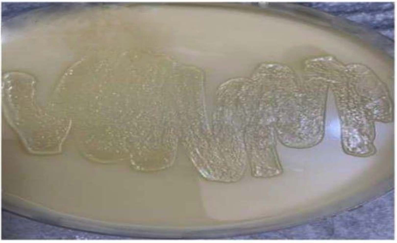
Figure 1. Growth of Lactobacillus isolate on MRS agar
ZnO NP biosynthesis by Lactobacillus
Several studies have explored the synthesis of ZnO NPs using various biological components. After synthesis, the reaction mixture appeared whitish and turbid, with NPs eventually settling at the bottom of the flask after several hours.25-27 The formation of a white pellet, as shown in Figure 2, served as visual confirmation of ZnO NP production. The collected material was placed on a small plate and dried in an oven at 40 °C for 8 hrs, yielding powdered ZnO NPs. These results are consistent with previous studies using Lactobacillus for ZnO NP synthesis28,29 and align with reported effects observed in similar research.30
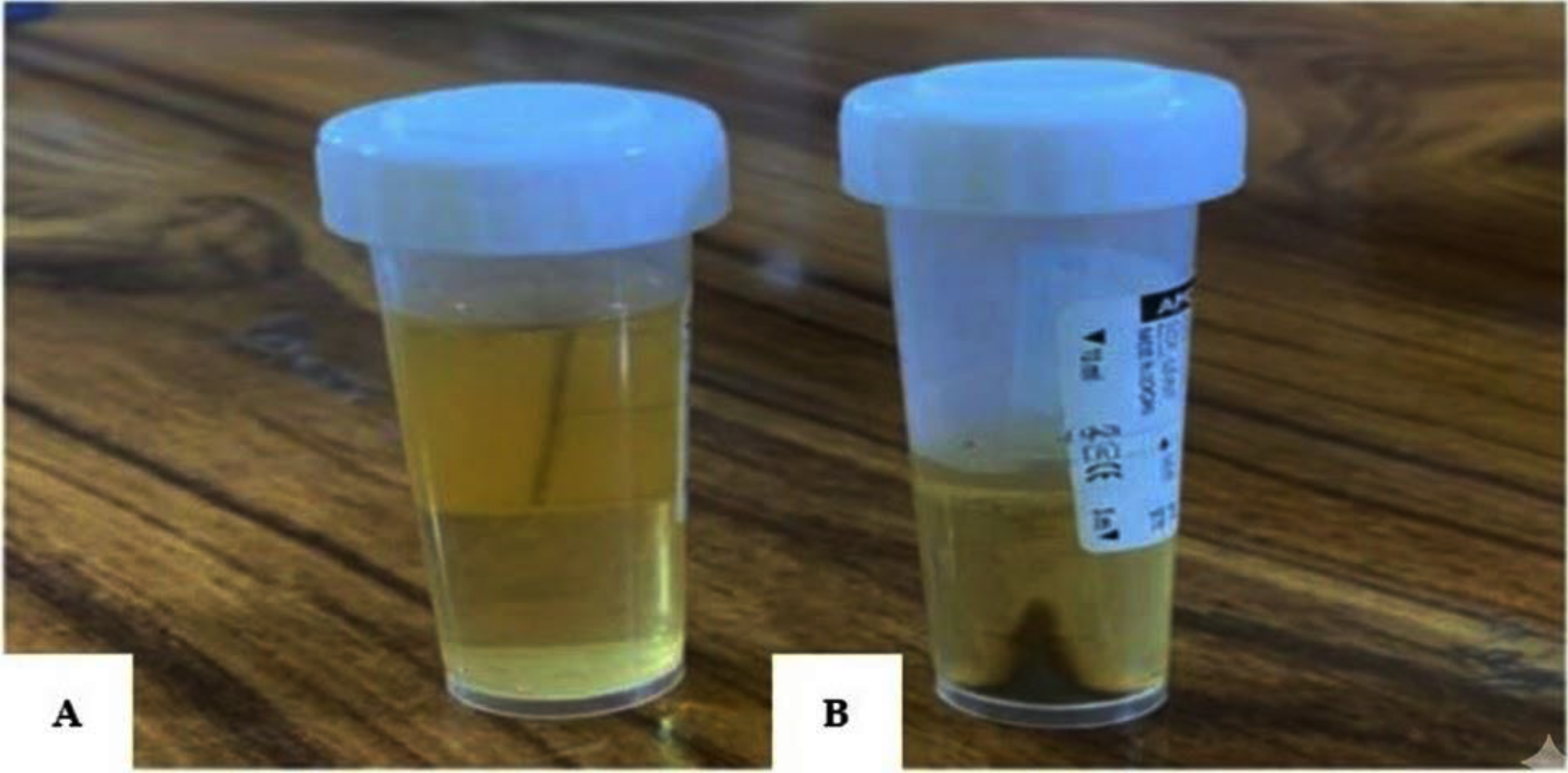
Figure 2. Biomanufacture of ZnO NPs using Lactobacillus spp. (A) Within 1 hr of adding zinc salts to the Lactobacillus cell-free supernatant. (B) After 72 hrs of incubation with zinc salts in the Lactobacillus cell-free supernatant
Analysis of ZnO NPs Using FE-SEM
FE-SEM was used to examine the morphology and size of ZnO NPs biosynthesized by Lactobacillus spp. After synthesis, the NPs were calcined at 500 °C for 1 hrs. Figures 3 and 4 show the FE-SEM images of the ZnO NPs. The images reveal that the NPs are hexagonal in shape, with diameters ranging from 56.14-64.97 nm, consistent with their classification as ZnO NPs.31
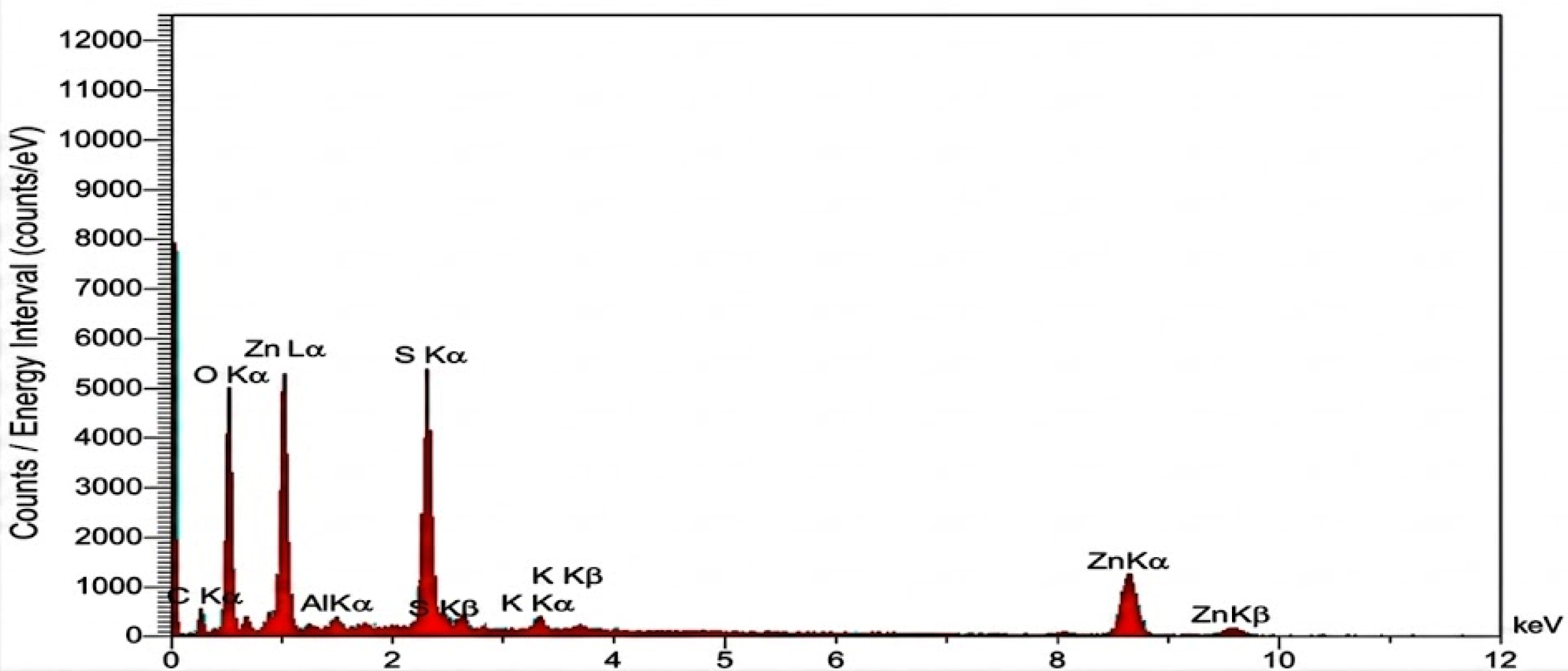
Figure 3. FE-SEM for ZnO nanoparticles
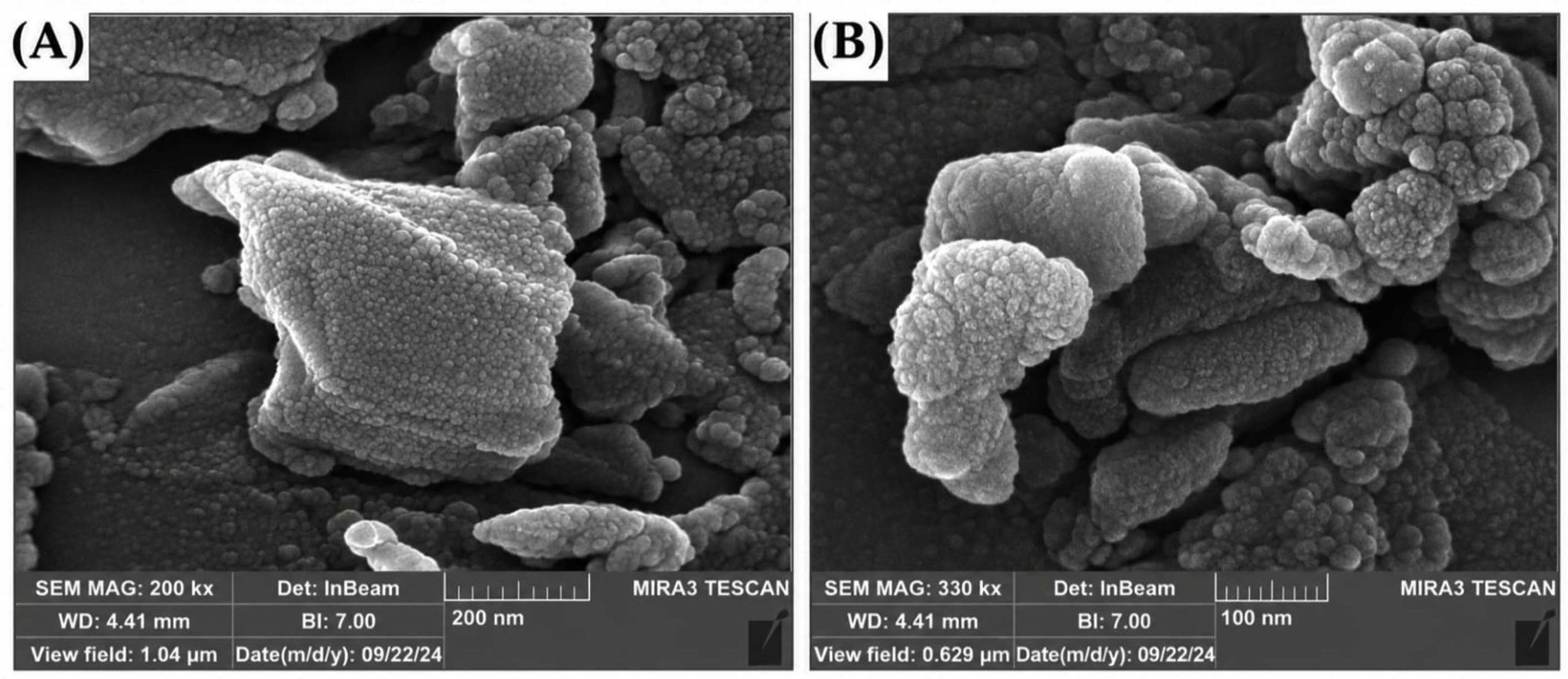
Figure 4. SEM micrographs of the prepared sample at different magnifications: (A) SEM image recorded at 200 k× magnification (scale bar = 200 nm), showing the platelet-like morphology of the particles; (B) SEM image recorded at 330 k× magnification (scale bar = 100 nm), showing the surface morphology and particle aggregation in greater detail
FTIR analysis
The functional groups involved in the synthesis of ZnO NPs were analyzed using FTIR spectroscopy. It is suggested that interactions between bacterial biomolecules and functional groups on bacterial cells contribute to the formation of ZnO NPs.27 Similarly, Hu et al. reported that functional groups derived from proteins, including -NH2, -OH, and -COOH, act as binding sites that facilitate zinc reduction (Figure 5).32,33
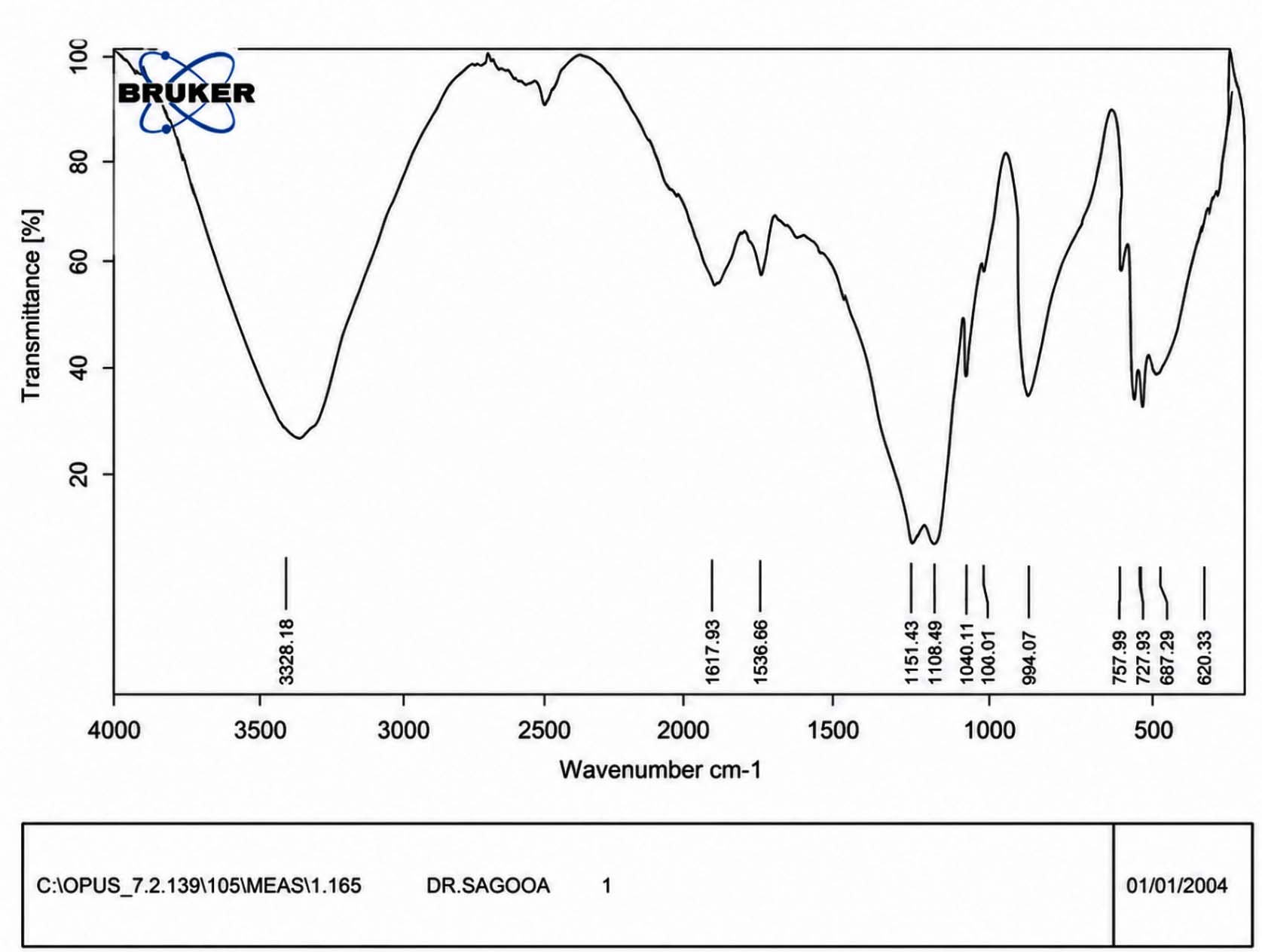
Figure 5. FTIR measurements for ZnO NPs
UV-Vis diffuse reflectance
The interference that affected the UV/Vis absorbance of the colloidal ZnO NPs at 257 nm was eliminated when the biomass was extracted from the samples. At first, centrifugation of the samples was considered a useful step. Nevertheless, the solution turned yellow at 257 nm, and the absorbance of the colloidal NP suspension dropped dramatically to nearly zero. This could be explained by the NPs adhering to each other or becoming entangled in the biomass. Therefore, centrifugation should be avoided, as it can destroy the NPs. The absorbance of the NPs was measured at 257 nm was measured and corrected before starting further operations. UV spectroscopy was used to track the colloidal ZnO NP solution in the 300-700 nm range (Figure 6).
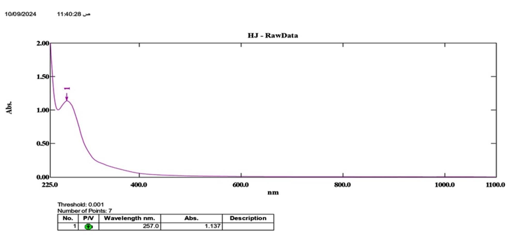
Figure 6. UV-Vis spectroscopic analysis of ZnO NPs
Antifungal Activity
The ZnO NPs demonstrated remarkable antifungal activity against the tested fungi, resulting in inhibitory zones ranging from 28-29 mm in diameter (Figures 7 and 8). In comparison, the antibiotic controls showed much smaller inhibitory effects, with fluconazole and ketoconazole generating zones of only 6 mm.34-36
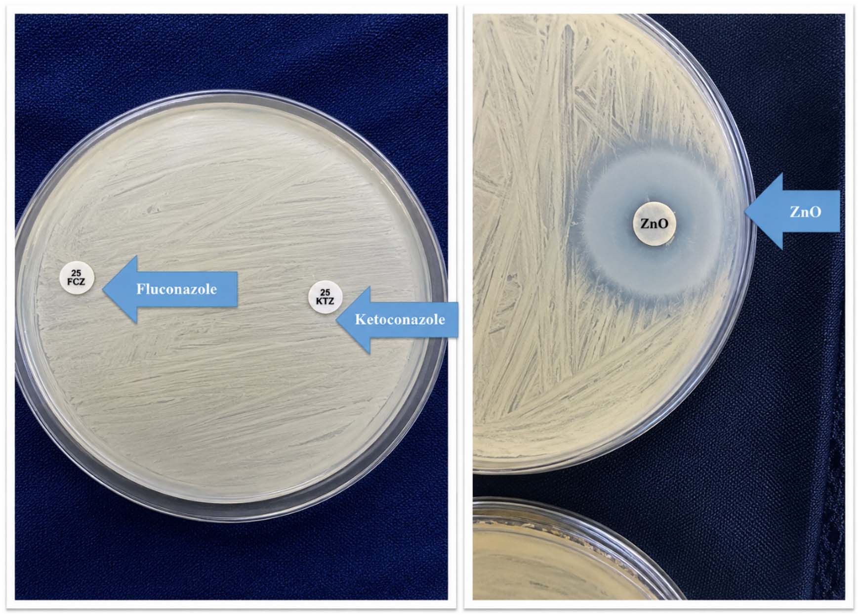
Figure 7. Comparison of antifungal activity between ZnO NPs and antibiotics against Malassezia globosa
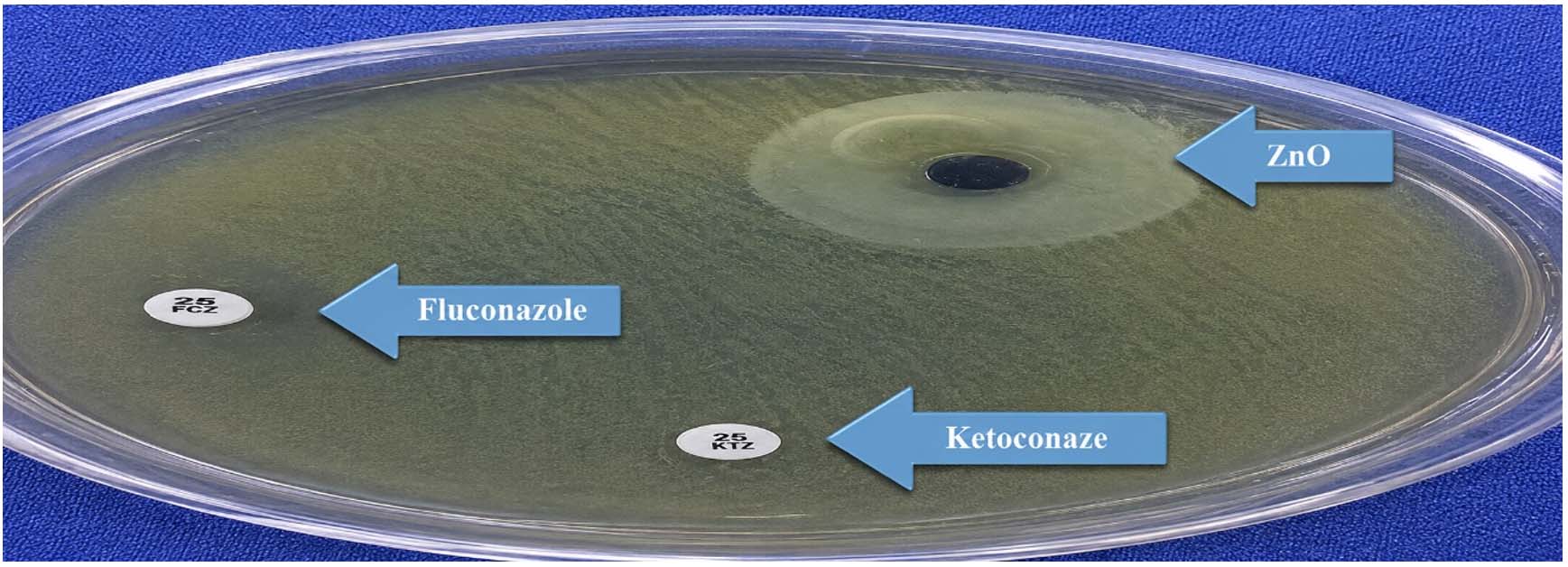
Figure 8. Comparison of antifungal activity between ZnO NPs and antibiotics against Candida albicans
Biosynthesized ZnO-NPs are considered to have significant growth-stimulating potential, in addition to their antimicrobial properties. In this study, a strain of Lactobacillus was isolated and identified using several characterization techniques for its ability to produce ZnO NPs. The results of multiple characterization procedures confirmed the successful synthesis of ZnO NPs with a spherical morphology and average diameters ranging from 56.14-64.97 nm. Furthermore, ZnO-NPs demonstrated strong antifungal activity against C. albicans and M. globosa, the latter being associated with dandruff on the scalp. These findings suggest that biosynthesized ZnO NPs could serve as a promising alternative for the treatment of various fungal infections. However, additional investigation is required to validate this hypothesis.
ACKNOWLEDGMENTS
The authors express their gratitude to the Department of Pharmacognosy, University of Thi-Qar, Thi-Qar, Iraq, for their valuable support.
CONFLICT OF INTEREST
The authors declare that there is no conflict of interest.
AUTHORS’ CONTRIBUTION
SHS and AHF conceptualized the study. HJM and HSY collected resources. MHM, JHA and AJH performed the experiments. BAG supervised the study. HKA wrote the manuscript. HKA and AHK reviewed and revised the manuscript. All authors read and approved the final manuscript for publication.
FUNDING
None.
DATA AVAILABILITY
All datasets generated or analyzed during this study are included in the manuscript.
ETHICS STATEMENT
Not applicable.
- Podagatlapalli GK. The fundamentals of synthesis of the nanomaterials, properties, and emphasis on laser ablation in liquids: a brief review. Discov Nano. 2025;20(1):98.
Crossref - Khan K, Javed S. Silver nanoparticles synthesized using leaf extract of Azadirachta indica exhibit enhanced antimicrobial efficacy than the chemically synthesized nanoparticles: A comparative study. Sci Prog. 2021;104(2):368504211012159.
Crossref - Rasmussen JW, Martinez E, Louka P, Wingett DG. Zinc oxide nanoparticles for selective destruction of tumor cells and potential for drug delivery applications. Expert Opin Drug Deliv. 2010;7(9):1063-1077.
Crossref - Huq MA, Ashrafudoulla M, Rahman MM, Balusamy SR, Akter S. Green Synthesis and Potential Antibacterial Applications of Bioactive Silver Nanoparticles: A Review. Polymers. 2022;14(4):742.
Crossref - Motelica L, Ficai D, Oprea O, et al. Biodegradable Alginate Films with ZnO Nanoparticles and Citronella Essential Oil: A Novel Antimicrobial Structure. Pharmaceutics. 2021;13(7):1020.
Crossref - Hossain A, Abdallah Y, Ali MA, et al. Lemon-Fruit-Based Green Synthesis of Zinc Oxide Nanoparticles and Titanium Dioxide Nanoparticles against Soft Rot Bacterial Pathogen Dickeya dadantii. Biomolecules. 2019;9(12):863.
Crossref - Mohd Yusof H, Abdul Rahman N, Mohamad R, Zaidan UH, Samsudin AA. Biosynthesis of zinc oxide nanoparticles by cell-biomass and supernatant of Lactobacillus plantarum TA4 and its antibacterial and biocompatibility properties. Sci Rep. 2020;10(1):19996.
Crossref - Singh H, Desimone MF, Pandya S, et al. Revisiting the Green Synthesis of Nanoparticles: Uncovering Influences of Plant Extracts as Reducing Agents for Enhanced Synthesis Efficiency and Its Biomedical Applications. Int J Nanomedicine. 2023;18:4727-4750.
Crossref - Anjum S, Hashim M, Malik SA, et al. Recent Advances in Zinc Oxide Nanoparticles (ZnO NPs) for Cancer Diagnosis, Target Drug Delivery, and Treatment. Cancers. 2021;13(18):4570.
Crossref - Kalaba MH, Moghannem SA, El-Hawary AS, Radwan AA, Sharaf MH, Shaban AS. Green Synthesized ZnO Nanoparticles Mediated by Streptomyces plicatus: Characterizations, Antimicrobial and Nematicidal Activities and Cytogenetic Effects. Plants (Basel). 2021;10(9):1760.
Crossref - Yadav S, Rani N, Saini K. Coupling ZnO with CuO for efficient organic pollutant removal. Environ Sci Pollut Res Int. 2023;30(28):71984-72008.
Crossref - Alkufi H, J. Kassab HJ. In-vivo examination of nasal-to-brain administration for delivering rhodamine B nanospanlastic for fluorescent vesicle imaging. J Res Pharm. 2026;30(2):569-575.
Crossref - Vijayakumar G, Kim HJ, Rangarajulu SK. In Vitro Antibacterial and Wound Healing Activities Evoked by Silver Nanoparticles Synthesized through Probiotic Bacteria. Antibiotics. 2023;12(1):141.
Crossref - Salman AH, Alkufi HK, Taher SS, Al-mahmood S, Haiss MA. Response surface optimization and in vitro study of nasal solusomes nanovesicles for the bioavailability improvement and brain targetting of sumatriptan. Epitheorese Klin Farmakol Farmakokinet. 2024;42(Sup1):107-117.
Crossref - Revankar AG, Bagewadi ZK, Aljaezi I, et al. Optimized collagenase biosynthesis (Bacillus siamensis strain Z1) and its application in collagen hydrolysate-mediated silver and zinc oxide nanoparticles synthesis and characterization with antibacterial, antioxidant and cytotoxic activities. PLoS One. 2026;21(3):e0344482.
Crossref - Chapot-Chartier MP, Kulakauskas S. Cell wall structure and function in lactic acid bacteria. Microb Cell Fact. 2014;13 Suppl 1(Suppl 1):S9.
Crossref - Sklodowski K, Chmielewska-Deptula SJ, Piktel E, Wolak P, Wollny T, Bucki R. Metallic Nanosystems in the Development of Antimicrobial Strategies with High Antimicrobial Activity and High Biocompatibility. Int J Mol Sci. 2023;24(3):2104.
Crossref - Mohammed YHI, Shntaif AH, Mansour AA, et al. Synthesis and multifaceted evaluation of novel AgCZ nanocomposite for targeted anti-angiogenic cancer therapy. Sci Rep. 2024;14(1):29289.
Crossref - Abd Sulaiman A, Kadhim Abbas H, Al-Samydai AM, Alkufi HK, Abdul Hadi Kharaba H, Al-hussaniy HA. A comparative diagnostic study for using the contrast agent in active and non-active multiple sclerosis by region of interest parameter. Biomed Pharmacol J. 2023;16(4):2531-2537.
Crossref - Durazzo A, Nazhand A, Lucarini M, et al. An Updated Overview on Nanonutraceuticals: Focus on Nanoprebiotics and Nanoprobiotics. Int J Mol Sci. 2020;21(7):2285.
Crossref - Arsalan N, Hassan Kashi E, Hasan A, et al. Exploring the Interaction of Cobalt Oxide Nanoparticles with Albumin, Leukemia Cancer Cells and Pathogenic Bacteria by Multispectroscopic, Docking, Cellular and Antibacterial Approaches. Int J Nanomedicine. 2020;15:4607-4623.
Crossref - Mazilu C, Deju R, Georgescu DP, Apostu A, Barbu A. Effects of Micro- and Nanosilica on the Mechanical and Microstructural Characteristics of Some Special Mortars Made with Recycled Concrete Aggregates. Materials. 2024;17(12):2791.
Crossref - Jain D, Shivani, Bhojiya AA, et al. Microbial Fabrication of Zinc Oxide Nanoparticles and Evaluation of Their Antimicrobial and Photocatalytic Properties. Front Chem. 2020;8:778.
Crossref - Humphries R, Bobenchik AM, Hindler JA, Schuetz AN. Overview of Changes to the Clinical and Laboratory Standards Institute Performance Standards for Antimicrobial Susceptibility Testing, M100, 31st Edition. J Clin Microbiol. 2021;59(12):e0021321.
Crossref - Khan F, Kang MG, Jo DM, et al. Phloroglucinol-Gold and -Zinc Oxide Nanoparticles: Antibiofilm and Antivirulence Activities towards Pseudomonas aeruginosa PAO1. Mar Drugs. 2021;19(11):601.
Crossref - Ghaffar N, Javad S, Farrukh MA, et al. Metal nanoparticles assisted revival of Streptomycin against MDRS Staphylococcus aureus. PLoS One. 2022;17(3):e0264588.
Crossref - Al-Tameemi AI, Masarudin MJ, Rahim RA, et al. Eco-friendly zinc oxide nanoparticle biosynthesis powered by probiotic bacteria. Appl Microbiol Biotechnol. 2025;109(1):32.
Crossref - Mohd Yusof H, Mohamad R, Zaidan UH, Rahman NA. Sustainable microbial cell nanofactory for zinc oxide nanoparticles production by zinc-tolerant probiotic Lactobacillus plantarum strain TA4. Microb Cell Fact. 2020;19(1):10.
Crossref - Asif N, Amir M, Fatma T. Recent advances in the synthesis, characterization and biomedical applications of zinc oxide nanoparticles. Bioprocess Biosyst Eng. 2023;46(10):1377-1398.
Crossref - Shamsuzzaman, Ali A, Mohd A, Mashrai A, Khanam H. Green synthesis of ZnO nanoparticles using bacillus subtilis and their catalytic performance in the one-pot synthesis of steroidal thiophenes. Eur Chem Bull. 2014;3:939-945.
Crossref - Luhar I, Luhar S, Al Bakri Abdullah MM, et al. Solidification/Stabilization Technology for Radioactive Wastes Using Cement: An Appraisal. Materials. 2023;16(3):954.
Crossref - Al-Askar AA, Hashem AH, Elhussieny NI, Saied E. Green Biosynthesis of Zinc Oxide Nanoparticles Using Pluchea indica Leaf Extract: Antimicrobial and Photocatalytic Activities. Molecules. 2023;28(12):4679.
Crossref - Hu P, Hu P, Vu TD, et al. Vanadium Oxide: Phase Diagrams, Structures, Synthesis, and Applications. Chem Rev. 2023;123(8):4353-4415.
Crossref - Mutukwa D, Taziwa RT, Tichapondwa SM, Khotseng L. Optimisation, Synthesis, and Characterisation of ZnO Nanoparticles Using Leonotis ocymifolia (L. ocymifolia) Leaf Extracts for Antibacterial and Photodegradation Applications. Int J Mol Sci. 2024;25(21):11621.
Crossref - Yassin MT, Mohamed S, Al-Otibi FO, Maniah K, AbdelGawwad MR. Synergistic antifungal activity of Lepidium sativum ZnO nanoparticles and nystatin against resistant Candida species. Sci Rep. 2025;15(1):34650.
Crossref - Xuan L, Ju Z, Skonieczna M, Zhou PK, Huang R. Nanoparticles-induced potential toxicity on human health: Applications, toxicity mechanisms, and evaluation models. MedComm. 2023;4(4):e327.
Crossref
© The Author(s) 2026. Open Access. This article is distributed under the terms of the Creative Commons Attribution 4.0 International License which permits unrestricted use, sharing, distribution, and reproduction in any medium, provided you give appropriate credit to the original author(s) and the source, provide a link to the Creative Commons license, and indicate if changes were made.