


ISSN: 0973-7510
E-ISSN: 2581-690X
Streptomycetes are considered as the most prolific microbes for screening of secondary metabolites like antibiotics for combating the situation of clinical infections. Presently, soil samples were collected from eight different locations of the rice field of West Godavari district, Andhra Pradesh. The collected soil sample (1 g) was taken and serial dilution was carried out to screen actinomycetes primarily over the different culture media like oatmeal agar, ISP1, and Starch Casein Agar and colonies appeared whitish-gray, greyish-white, and turmeric-yellow. For the antimicrobial activity these microbes were tested over the Gram-positive (Bacillus subtilis), Gram-negative bacterium (E. coli) and fungi Aspergilus niger. Positive results reported in bacterium and negative against fungi. Extraction of antibiotics was done in conical flask using Yeast Extract-Malt Extract broth and inoculated with screened overnight starter culture and put it for fermentation at 28 °C for 8 days in rotary shaker and after completion supernatant was collected by centrifugation and crude collected by vortexing with equal ethyl acetate. 16S rRNA sequencing was done to identify the potential microbes and reported as Streptomyces avermitilis strain ZRS13. Antimicrobial activity of crude extract were reported through cell wall damage observed via FESEM using FESEM and PI staining. Here the present study concludes that rice field of Tadepalligudem are an abundant source of Actinomycetes, Streptomyces avermitilis ZRS13 which is capable to inhibit Bacillus subtilis and E. coli but didn’t showed any antimicrobial activity against Aspergillus niger. Antimicrobial activity were purported by mechanism like damage of bacterial cell wall, confirmed by FESEM and PI staining. From this study it confirmed that Streptomyces avermitilis ZRS13 can be used as potent strain for developing new antibiotics.
Streptomycetes, Antibiotics, Secondary Metabolites, FESEM
Microorganisms have long been considered as an abundant source of natural products for both industrial and medical purpose. Microorganisms isolated from soil have widely been exploited for the screening of many primary and secondary metabolites. Among these soil bacterium Actinomycetes are recognized as an ultimate source for many enzymes and antimicrobials. Within Actinobacteria most dominant genus studied and found to have highs is Streptomyces which are referred as the biological antagonist.1 These Streptomyces are filamentous, spore-forming, and Gram-positive within phylum Actinobacteria.2,3 Throughout the golden age of antibiotic discovery, Streptomyces have been used for their ability to produce bioactive metabolites.4
These are alone enough to provide more than 70% of the total antibiotics having commercial value and still being screened for novel antibiotics against antibiotics resistant pathogenic microbes.5 These are usually a Gram-positive filamentous bacteria that belongs to diverse groups basically reported from various sites including soils, fresh water sites and many extreme or unexplored area could be major sites of Streptomyces.6 About 850 Streptomyces are well studied and found to have high G+C% (≥70%) along with large genome size of 8-10 Mb.7
As these Streptomyces are considered as the most abundant reservoir for natural products due to their complex secondary metabolism.8 Streptomyces are capable to produce around 10000+ secondary metabolite especially antibiotics for clinical and other agricultural based purpose.9 Major antibiotics produced by the Streptomyces are mainly including macrolides, tetracyclines, aminoglycosides, glycopeptides, ansamycins, and terpenes. Alone, Streptomyces hygroscopicus is capable to produce 180 types of secondary metabolites having wide antimicrobial activity. With having such wide potential to produce various secondary metabolite specially antibiotics against the pathogenic microbes the chance of antimicrobial resistance is increasing abruptly which can cause mortality up to 10 million per year with an economic burden.10 It claims over millions of lives over globe annually, which includes children and aged citizens most vulnerable.11 Multidrug-resistant bacteria (MDR), colloquially termed as “superbugs” make the situation even more pathetic. Few decades ago, the emergence of multidrug-resistant bacteria increased devastatingly. The most common multidrug-resistant bacteria are Escherichia coli resistant against cephalosporin and fluoroquinolones, Klebsiella pneumonia against cephalosporin and carbapenems, Staphylococcus aureus to methicillin and vancomycin, Neisseria gonorrhoeae to cephalosporin. Making possible combination of ineffective drugs against multidrugs-resistant microbes with some adjuvants showed hope to keep desired effects.12 Novel metabolites screened especially from Streptomyces, offers a significant opportunity for treatment of disease with efficiency, to minimize this kinds of terrible consequences with notable solution.
Collection of soil samples
Eight soil samples were collected from different random location of rice fields nearby village of Tadepalligudem, Andhra Pradesh, India. Using trowel of normal size, soil sculpt at a depth of about 10 cm and diameter of 5 cm soil samples were collected from the rice-fields. Collecting soil sample from rice fields transferred directly to sterile polythene bags and carried directly to Microbiology laboratory. Collected soil samples were kept at stored at 4 °C (refrigeration) inside the refrigerator till further analysis. The soil sample collection from various sites were done with proper mentioning of GPS location given in Table 1.
Table (1): Cultural characteristics of Actinomycetes grown over various culture media
No. |
GPS Location |
Soil appearance |
Sampling depth (cm) |
Name of culture media |
Colonies Color |
Growth |
|---|---|---|---|---|---|---|
01 |
16°50’09.9″N 81°31’29.2″E |
Red soil |
10 |
Casein Starch agar |
Pink |
++ |
02 |
16°50’07.7″N 81°31’25.9″E |
Muddy soil |
10 |
Nutrient agar |
White |
+ |
03 |
16°50’07.7″N 81°31’33.5″E |
Red soil |
10 |
ISP1 |
Grey |
+++ |
04 |
16°49’48.4″N 81°31’43.3″E |
Muddy soil |
10 |
ISP2 |
White |
+++ |
05 |
16°49’48.4″N 81°31’54.3″E |
Browny earth |
10 |
ISP3 |
Grey |
++ |
06 |
16°49’48.4″N 81°31’63.3″E |
Red soil |
10 |
ISP4 |
Yellow |
++ |
07 |
16°50’07.7″N 81°31’34.9″E |
Red soil |
10 |
PDA |
White |
+ |
08 |
16°50’09.9″N 81°31’49.2″E |
Red soil |
10 |
Oatmeal agar |
Black |
++ |
+++: Good activity; ++: Moderate activity; +: Weak activity
Isolation of Actinomycetes/Streptomycetes
Soil samples collected from different locations were serially diluted (10-4) dilution using one gram of soil dissolved in sterile water and inoculated on ISP-2 media.13 Penicillin (20 mg/L) were added to the medium to retard the growth of bacteria and fungi, likewise. Later on all the plates were incubated at 30 °C ± 2 °C for 5-7 days. Colonies isolated from soil samples were white, grey, yellow, pink and black pigmented, respectively (Figure 1). Interestingly, gray and white mycelial-pigmented Actinomycetes were prominent in the soil. Pure culture streaked on ISP-2 agar plate and stored at 4 °C inside the refrigerator till further use. The cultural characteristics of potent Streptomycetes over different culture media is given in Table 1.
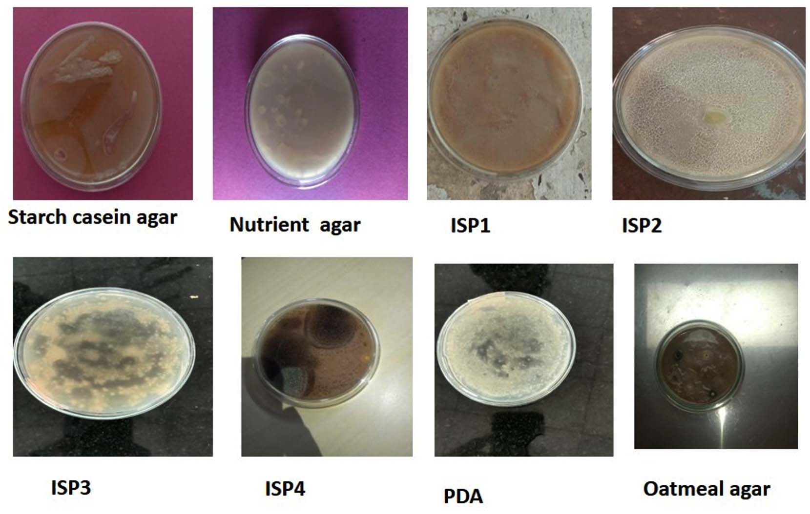
Figure 1. Screening of microbial isolates with various media
Morphological studies
Culture plates with Actinomycetes were prepared and covered with cover-slips and placed at an inclined angle of 45 °C and incubated at 28 °C for 7-8 days. Later on cover slips were removed carefully and examined under the microscope. Conidiospores on located on aerial and substratum mycelia and was observed and compared with the Bergeys Manual of Determinative Bacteriology.
Primary screening of potent microbes from the culture plates
For screening of potent microbes against the pathogenic microbes were done by culturing it on the agar plates. The pathogenic strains were spread over the agar plates and after few minutes of drying the potent microbes were streaked from the culture plates as shown in Figure 1. The selected microbes were streaked over the lawn of the bacterial and fungal culture on plates and placed inside the incubator at 37 °C overnight. The antimicrobial activity of selected strains were noticed on the next day (Figure 1). The screening of antimicrobial activity of selected Actinomycetes are given in Table 2. Among the selected strains were again tested for antimicrobial activity against bacterium is shown in Figure 2.
Table (2): Screening of antimicrobial activity of Actinomycetes isolates
No. |
Isolates code |
Bacillus subtilis |
E. coli |
Aspergillus niger |
|---|---|---|---|---|
1 |
Stp1 |
– |
+ |
– |
2 |
Stp2 |
++ |
+++ |
– |
3 |
Stp3 |
+ |
– |
– |
4 |
Stp4 |
– |
– |
– |
5 |
Stp5 |
+ |
– |
– |
6 |
Stp6 |
– |
+ |
– |
7 |
Stp7 |
++ |
– |
– |
8 |
Stp8 |
– |
+ |
– |
+++: Good activity; ++: Moderate activity; +: Weak activity; -: No activity
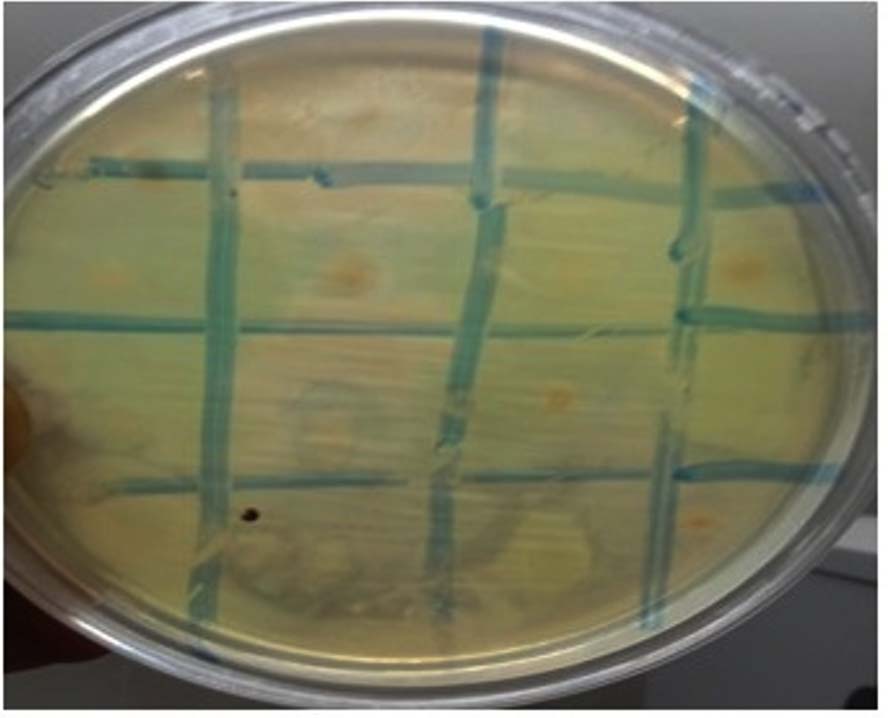
Figure 2. Primary Screening of selected strains for antimicrobial activity against the bacteria
16S rRNA sequencing and phylogenetic analysis
Identifying the isolated species taxonomical nomenclature given by molecular methods, 16S rRNA molecular analysis was performed (Figure 3). 16S rRNA sequence and Blast analysis provide identification of the isolated species. The 16S rRNA sequencing were outsourced from BioEdge solution, Bangalore, Karnataka, India and their protocol mentioned in text with details and likewise their protocol also described.
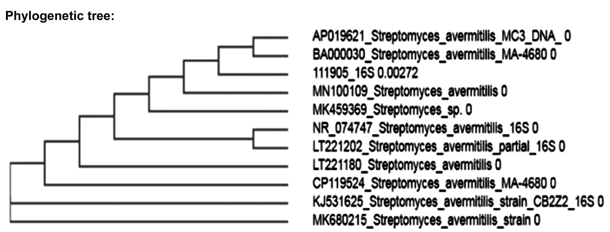
Figure 3. Phylogenetic tree of Streptomyces avermitilis strain ZRS13 microbial isolates with various media
Microbial culture
To screen out the Actinomycetes against microorganism different strains of bacteria including E. coli (Gram-negative) and Bacillus subtilis (Gram-positive) were taken. In case of Fungi Aspergillus niger were taken for primary screening. The tested microbes were taken from Department of Biotechnology, NIT Andhra Pradesh, Tadepalligudem, Dist.-West Godavari, Andhra Pradesh, India.
Crude antimicrobial metabolite extraction and antimicrobial activity/Secondary screening
For extracting the secondary metabolites was conducted by submerged fermentation. A 24 hours old an inoculum (seed culture) of 100 ml was used to incubate 500 ml of Erlenmeyer flask of Yeast Extract-Malt Extract broth was incubated at 30 ± 2 °C on rotary shaker for 8 days.
Fermentation comes to halted on 8th days, broth was filtered through Whatman No. 1 filter paper. 70% ammonium sulphate was used to precipitate out proteinaceous materials from fermentation broth. The suspension obtained was centrifuged at 4000 rpm for 20 minutes to settle down other debris particles. An equal amount of ethyl acetate (HiMedia, Cat. Number: 141-78-6) was added to the centrifuged suspension and vortexed for 1 hour. A separating funnel was used to separate the aqueous and organic phases for 5 min. The upper organic phase was evaporated at a 40 °C water bath, and a small amount of pH 4 phosphate buffer was added to solubilize crude antibiotic extract.
Determination of antimicrobial activity
Using sterile cork borer 6 mm diameter of agar wells were prepared and loaded with crude extract of 800 µg/mL, negative control (PBS) were used. The plates were incubated at 24 hrs at 37 °C for antibacterial activity.13 Likewise, potato dextrose agar and Mueller-Hinton agar for fungi and bacteria were poured into the sterilized Petri plates for well diffusion assay for determining the anti fungal and antibacterial activity. Using sterilized spreader 1 ml of the sample culture spread over the PDA plates. Agar wells of 6 mm diameter was prepared and loaded with crude extract concentration of 800 µg/mL, using PBS as negative control and incubated for 48 hours for assessing the anti-fungal and antibacterial activity.
Field emission scanning electron microscope
For detailed analysis of the change in the morphology of the bacterial cell field emission scanning electron microscopy was used. Bacteria were grown in the Nutrient broth and treated with the defined concentration for 24 and 48 hours. To settle down the bacterial pellet centrifuged at 6000 rpm for 10 minutes. Settled bacterial pellets were washed twice a time with phosphate buffer saline (PBS) at 5000 rpm for 5 minutes and later fixed with 2.5% glutaraldehyde solution for 1 hr. Pellets were fixed with glutaraldehyde solution, again washed with Phosphate buffer saline (PBS) at 5000 rpm for 5 minutes and subjected for dehydration in a series of 10, 20, 30, 40, 50, 60, 70, 80, 90 and 100% ethanol solution for 10 minutes at 37 °C. Dehydrated bacterial pellet were finally put inside the incubator for complete drying at 65 °C for the next 3 days respectively. Completely dried bacterial pellet were then further coated with Palladium:Gold (80:20) for field emission scanning electron microscopy (Figure 4). Sample preparation for the field emission scanning electron microscopy was done using standard protocol with minor modifications.14
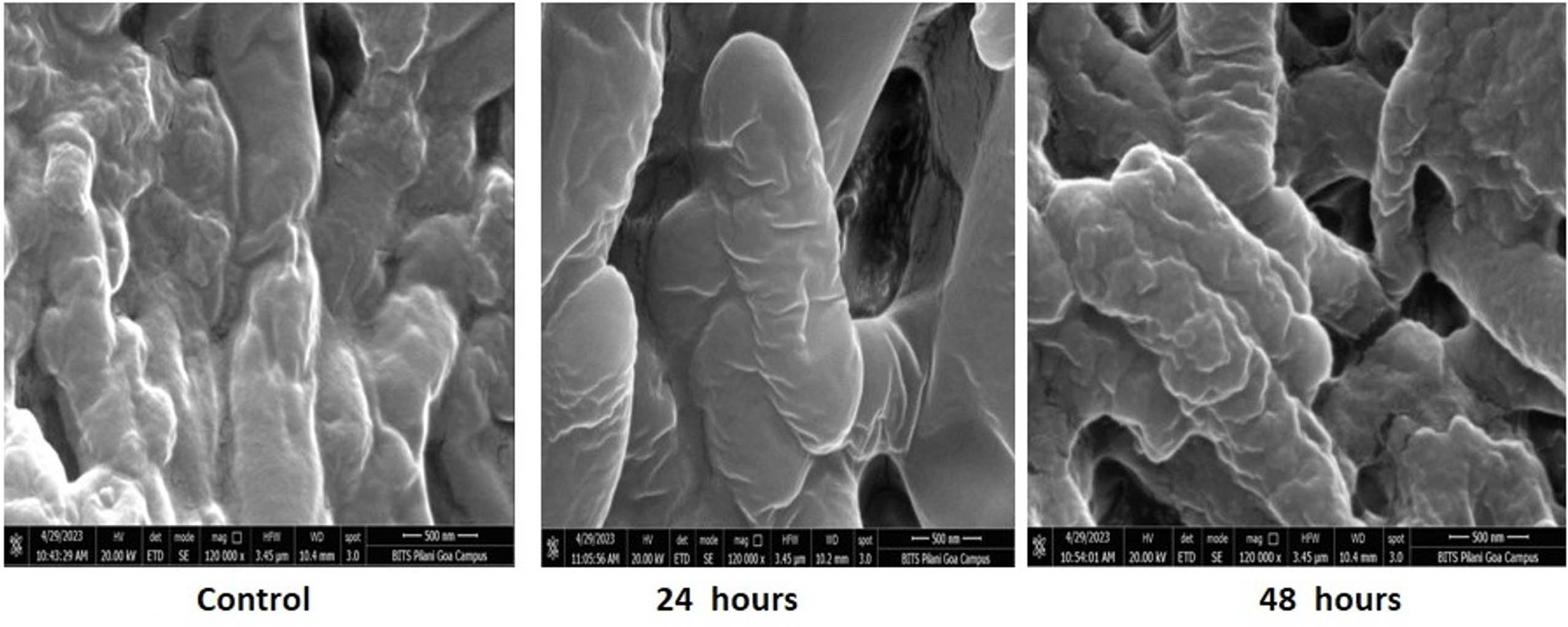
Figure 4. Field Emission Scanning Electron microscopy microbial isolates with various media
Cell viability test
The bacterial cell were tested against crude extract for various time intervals viz. 24, 48 and 72 hours respectively. The viability was judged by using fluorescent microscopy by staining cells with Propidium iodide (PI) at 20× magnifications. Propidium iodide is commonly known as dead cell marker as it bind to bio-molecule DNA when the cell membrane of dead cell is damaged. When bacterial cells are stained with Propidium iodide red fluorescence appears which indicates that cell is dead and it binds to bio-molecule DNA
(Figure 5).
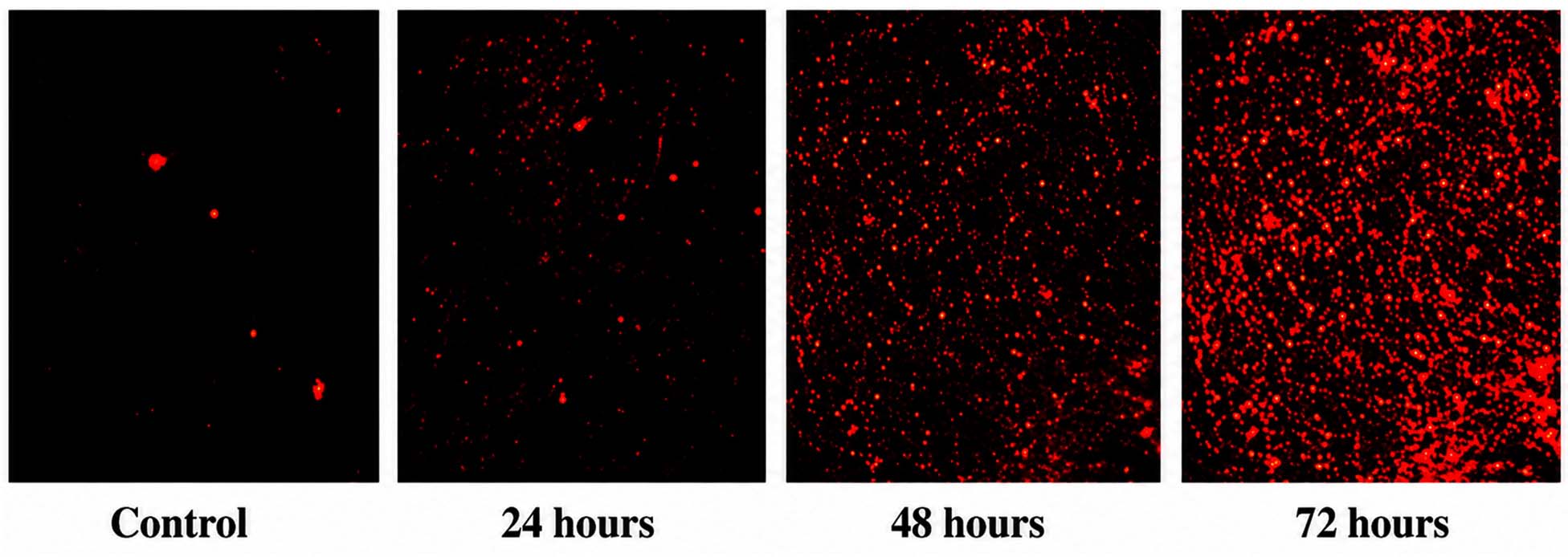
Figure 5. (a) PI (Propidium Iodide) Staining microbial isolates with various media
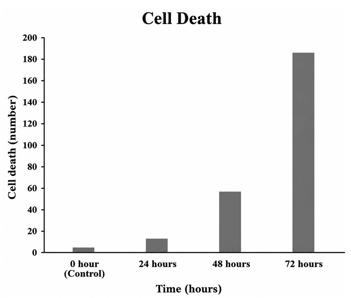
Figure 5. (b) Apoptotic cells after 24, 48 and 72 hours in Propidium Iodide cell staining microbial isolates with various media
Total 8 soil samples were collected from the different location of rice field of nearby village area of Tadepalligudem. Morphologically different actinomycetes colonies obtained after growing on different culture media were further screened by sub-culturing and stored at 4 °C to be used for further analysis. The isolated actinomycetes colonies spore color was categorized into white, grey, yellow, pink and black pigmented, respectively. Interestingly, gray and white mycelial-pigmented actinomycetes were prominent in the soil (Table 1).
Antimicrobial activity of the isolated strains
The antimicrobial activity of crude extract was determined by the well diffusion assay against the pathogenic microbes. Extract showed activity against the bacterial pathogens (E. coli and Bacillus subtilis) was found to be potent but not satisfactory against the fungal pathogens Aspergillus niger.
Extraction of crude antimicrobial compounds
Through centrifugation fermentation broth containing antimicrobial compounds were screened out from potent Streptomycetes. Crude obtained were tested for antimicrobial activity against microbes. The crude antimicrobial compound was tested against E. coli, Bacillus subtilis and Aspergillus niger but showed best potential activity against E. coli (Table 2).
Identification of selected strain
The Streptomyces isolate Stp2 with potent antibacterial activity was selected for the further characterization that based on morphological, physiological and molecular identification by 16S rRNA sequencing. The microscopic observation under the light microscope revealed that Stp12 form an unique pattern thread like structure. These morphological pattern later on further microscopic studies placed this isolates under the genera Streptomyces genera. Molecular identification of screened bacterium using 16S rRNA methods were done starting with CTAB method opted by outsourced firm. Here 500 µl of CTAB (Cetyl-trimethyl ammonium bromide) buffer was poured in bacterial pellet and simultaneously mixed and vortexed likewise. After proper mixing and vortex homogenate was transferred to 60 °C water bath for 30 minutes. Later on completion of incubation period, homogenate was centrifuged to 14000 × g for 5 minutes. Following centrifugation, equal volume of chloroform:Isoamyl alcohol (24:1) were added and vortexed for 5 minutes for proper mixing which followed by centrifugation at 14000× for 5 minutes to separate two layers likewise. After centrifugation the upper layer was collected and transferred to new tubes. Here DNA precipitation was done by adding 0.7 volume of chilled isopropanol and incubated at -20 °C overnight. Next day, 750 µl of DNA sample were transferred to DNA column and spun at 1200 rpm for 1 minutes. Following centrifugation now wash buffer (750 µl) were added and again spin at 12000 rpm for 1 minutes. This process is repeated for one more time. After completion of washing DNA columns were dried for 2 minutes at 12000 rpm. For better purification of isolated DNA sample 20 µl of elution buffer were added and kept at room temperature for 3 minutes following centrifugation 12000 rpm for 1 minutes. For mitigating RNA contamination from sample 1 µl of RNase enzyme was added with incubation of 37 °C for 30 minutes. The eluted DNA obtained were quantified using agarose gel electrophoresis shown in Figure 1.
Likewise running eluted DNA sample over agarose gel were later on subjected to PCR for amplification. PCR followed by using 27 F forward and 1492 R reverse primer. The sequence of these primers viz. 27 F forward primer (5′-AGAGTTTGATCCTGGCTCAG-3′) and 1492 R reverse primer (5′-GGTTACCTTGTTACGACTT-3′) with annealing temperature of 53 °C and 57 °C likewise. Here for PCR reaction containing 1 µl DNA template (25 ng), 2 µl 10× reaction buffer, 0.5 µl MgCl2 (50 pM), 1 µl dNTPs mix (10 mM), 1 µl forward primer (10 pM), 1 µl reverse primer (10 pM), 0.5 µl Taq polymerase (5 U/pi) and the final volume 25 µl adjusted with molecular grade water. Primers used here are basically standard primers available for 16S rRNA gene amplification. After preparing reaction mixture PCR conditions were adjusted. In PCR initial denaturation was conducted at 95 °C for 2 minutes and denaturation at 95 °C for 30 seconds only. Annealing at 50 °C for 30 seconds and elongation occurs at 72 °C for 1 minute. Denaturation, annealing and elongation steps were repeated for 30 cycles. In last final elongation took place at 72 °C for 10 minutes and held at 4 °C. After completion of PCR cycle PCR product were run over agarose gel, reported to be 1200 bp compared to 1000 bp ladder. Likewise eluted for purification, in last subjected for sanger sequencing following instruction. After sanger sequencing, post sequencing and PCR purification were done. After post sequencing data were obtained and analyzed by BioEdit software. The 16S rRNA sequencing analysis used for the BLAST analysis. 16S rRNA sequence was determined and Blast analysis was performed, which confirmed that the isolate belonged to the Streptomyces species (Bases 1-1428 linear RNA). The sequence data were analyzed by BLAST server and later on FASTA. The obtained sequences are given here below:
TTCACGGAGAGTTTGATCACTGCGTGATCT ATGAAGCCCTTCGGGGTGGATTAGTGGCGAACGGGTGAGTAA CACGTGGGCAATCTGCCCTGCACTCTGGGACAAGCCCTGGAAACGGGGTCTAATACCGG ATAATACTCTCGCAGGCATCTGTGAGGGT TAAAAGCTCCGGCGGTGCAG GATGAGCCCGCGGCCTATCAGCTTGTTGGTGAGGTAGTGGCTCAC CAAGGCGACGACGGGTAGCCGGC CTGAGAGGGCGACCGACTGA CACTGGGACTGAGACACGGCCCAGACTCCTACGGGAGGCAGCAGTGGGGAA TATTGCACAATGGGCGAAAGCCT GATGCA GCGACGCCGCGTGAGGGATGACGGCCTTCGGGTT GTAAACCTCTTTCAGCAGGGAAGAAGCGAAAGTGACGGTACCTGCAGAAGAAGCGCCGGCTAACTACGTGCCAGCAGCCGCGGTAATACGTA GGGCGCAAGCGTTGTCCGGAATTATT GGGCGTAAAGAGCTCGTAGGCGG CTTGTCACGTCGGGTGTGAAAGCCCGGG GCTTAACCCC GGGTCTGCATTCGATACGGGCTAGCTAGAGTGTGGTAGGGGAGATCGGA ATTCCTGGTGTAGCGGTGAAATGCGCAGATATCAGGAGGAACACCGGTGGCGAAGGCGG ATCTCTGGGCCATTACTGACGACGTAGCGACTAGCTAGTC GGTAGTCCACGCCGTAAACGGTGGGAACTAGGTGTTGGCGACATTCCAC GTCGTCGGTGCCGCAGCTAACGCATTAAGTT CCCCGCCTGGGGAGTACGGCCGCAAGGC TAAAACTCAAAGGAATTGACGGGGGCCCG CACAAGCAGC GGAGCATGTGGCTTAATTCGACGCAA CGCGAAGAACCTTACCAAGGCTTGACA TACACCGGAAAGCATTAGAGATAGTGCCCC CCTTGTGGTCGGTGTACAGGTGG TGCATGGCTGTCGTCAGCTCGTGTCGTG AGATGTTGGGTTAAGTCCCGCAACGAG CGCAACCCTTGTTCTGTGTTGCCAGCATGCCCTTC GGGGTGATGGGGACTCACAGGAGACCGCCGG GGTCAACTCGGAGGAAGGTGGGGACGA CGTCAAGTCATCATGCCCCTTATGTCTTGGG CTGCACAC GTGCTACAATGGCCGATACAATGAGCTG CGATACCGCAAGGTGGAGCGAATCTCAAAA AGTCGGTCTCAGACGAGCATCTACTCGCA GATCAGCATTG CTGCGGTGAATACGTTCCCGGGCCT
The identification of particular strain were conducted in outsourced way by BioEdge firm, Bangalore, Karnataka, India. The isolated strain has 97.53% similarity with Streptomyces sp. It is found that the strain is Streptomyces avermitilis strain ZRS13 with accession number MN100109.1 and A phylogenetic tree was constructed with bootstrap values in Figure 3.
Field emission scanning electron microscopy
The E. coli cells treated for 24, 48 hours respectively for the comparison of treated and untreated samples. The surface morphology of E. coli cells treated with crude extract of Streptomyces avermitilis strain ZRS13 showed significant shrinkage in the size of cells. At 500 nm After 24 hours of treatment cells seems smaller in size as compared to control. Furthermore it is also clearly visible that crude extract of isolated from Streptomyces avermitilis strain ZRS13 getting adhered over its surface topology. Similarly in case of 48 hours treatment showed that cells are being bends and cracks are quite visible in them. Furthermore it is also quite clear from the image at 500 nm that layering starts happening in them and cell wall starts degraded in layered pattern. The FESEM image clearly indicates that crude extract of Streptomyces avermitilis strain ZRS13 exhibiting quite significant antibacterial effect over E. coli. It also revealed that crude antimicrobial extract works in a time dependent manner.
Cell viability assay
Bacterium E. coli was subjected to crude antimicrobial extract of Streptomyces avermitilis strain ZRS 13 for the various time interval viz. 24, 48 and 72 hours respectively. Propidium iodide is mainly used as dead cell markers that binds directly to dead cell bio-molecule DNA after their cell wall getting ruptured. When E. coli cells stained with PI red fluorescence appear as dead cell acquired the stain and appear as red colour spot when observed by fluorescence microscope as given in Figure 5(a). From the result it is clearly shown that few Population of E. coli bacterium are acquiring stains of Propidium iodide in control; however E. coli population of smeared bacterial cells increased with increasing time interval at the same concentration.
Statistical analysis
For determination of antimicrobial activity of while screening of microbes from soil sample primary screening method were opted. After pouring of diluted soil sample over various microbial media and incubating for overnight, some potent bacterium showed zone of inhibition around themselves which confirms that microbes are releasing antimicrobial compounds which is retarding the growth of nearby bacterium. Likewise, those strains were then subjected for secondary screening and after fermentation their crude extract were used for determination of antimicrobial activity by well diffusion assay where they showed zone of inhibition. Yet the zone of inhibition was minor due to its impure nature but for qualitative data used other techniques also to measure cell count after apoptosis.
For determination of apoptosis in bacteria bacterial staining were done using propidium iodide whose function mainly based over binding of dye to the DNA molecules directly after cell wall breaking. The cell which undergoes apoptosis stained red in color their count were taken after every 24, 48 and 72 hours likewise and given in Figure 5(b). As much the cell appears red in color with time indicates cell death is happening with time. The counting of cells was done manually without using any kind of AI tools.
The present study was conducted for isolation of Streptomycetes from the soil samples collected from the various sites of rice fields of Tadepalligudem, Dist.-West Godavari, Andhra Pradesh, India. The main emphasize was on screening of Streptomyces spp., producing various secondary metabolites especially antibiotics for combating clinical infections and diseases.
A total of eight sites were selected from various rice fields of Tadepalligudem village for collection of soil samples for screening of Streptomyces spp. All strains were tested against microorganism including both Gram-positive bacteria (Bacillus subtilis), Gram-negative bacterium (E. coli) and Fungi Aspergillus niger likewise. Cultural characteristics of actinomycetes grown on various culture media and patterns of antimicrobial activity is shown in Tables 1 and 2, respectively. In the present study out of eight isolates showed their antibacterial activity but none showed its anti-fungal activity in primary screening.
The isolates obtained from the primary screening were observed under the microscopes and reported to have thread like structure, i.e. the main characteristics of Streptomyces for identifying on the morphological method. The present research reported that isolated strains having various color that makes them morphologically different starting from pink, white, gray, yellow, and black respectively. Streptomyces isolates are morphologically characterized mainly the formation of soluble pigment formation, characteristics of spores and color of aerial and substrate mycelium.15
In the current research actinomycetes isolates namely Streptomyces avermitilis strain ZRS13 was reported to have a spore chain with similar smooth surface, major characteristics of Streptomycete. The actinomycetes were grown on various media including Nutrient agar, oatmeals agar, Potato dextrose agar, ISP1, ISP2, ISP3, ISP4 and Casein starch agar. Out of these ISP1 and ISP2 media reported to have good growth of it.
For identification of Actinomycetes 16S rRNA sequencing played a major role in it as evident by many previous work.16,17 In this current study the molecular identification of Actinomycetes isolate Stp2 was reported Streptomyces sp. as Streptomyces avermitilis strain ZRS13 with accession number MN100109.
Antimicrobial compounds usually derived from plant origin, but presently it is also produced from many soil microbes including Actinomycetes; as they act as potent antibacterial agents.18 The present study reported that Streptomyces avermitilis strain ZRS13 is capable to produce antimicrobial compounds in Yeast Extract-Malt Extract broth (ISP-2). Similar kind of antimicrobial activity was reported by the marine Actinomycetes isolated from the East coast of India named Streptomyces afghaniensis VPTS3-1 against many different bacteria.19 Actinomycets isolated from the soil sample showed good antibacterial activity against both Gram-positive and Gram-negative bacterium.20 Some other studies where Actinomycetes isolated from Thar desert, India an arid zone region showed good antibacterial activity against various pathogenic bacteria by their purified components.21
Membrane integrity disruption leads to leakage of internal cellular content which is ultimately responsible for death of bacterial cells. The morphology of untreated bacterial cells were found to be smooth and even. The effect of certain herbal extract often possess the same effect like crude antimicrobial compound extracted from microbes.22,23 Primary targets of the β-lactam agents like Penicillin are penicillin binding proteins. The Penicillin binding proteins mainly interact with β-lactam ring and ceased the synthesis of new peptidoglycan.24
Propidium iodide is mainly used as the cell death marker and interact with the DNA of dead cell when the membrane potential is lost. Result here showed that after treating E. coli cells with crude antimicrobial extract for 24, 48 and 72 hours showed remarkable apoptosis in them.
Results are quite similar to the apoptotic effects reported by the various plant extracts over other microbes. The significant amount of increase in PI fluorescence intensity by exposing them with crude antimicrobial extract for 24, 48 and 72 hours shows increase in the loss of membrane integrity as the time interval increases with respect to time. Such presence of maximum number of cells stained with PI indicates maximum cell death in the given sample, which correlates with the data obtained from the PI.
The noticeable increase in PI fluorescence intensity after exposing them with the mixture for various time intervals viz. 24, 48 and 72 hrs shows the enhancement in loss of membrane integrity as the time interval increases with respect to time. The presence of maximum number of cells stained with PI fluorescence of FM as shown in Figure 5. Similarly, the PI fluorescence intensity is found to be increased at various concentrations after exposing Cr(VI) as compared to control showed that the enhancement in loss of membrane integrity is directly proportional to concentration of chromium.25
Actinomycetes are the microbes that are found in the different environments and are capable of producing novel antibiotics of industrial importance. In this present study total 8 samples were collected from the various sites of rice fields for screening of streptomycetes that are capable of producing secondary metabolites. The strains obtained after screening was reported to be Streptomyces avermitilis strain ZRS13 having antibacterial potential. Further, this study may be helpful to develop potential antibacterial and anti-fungal agents against different pathogens.
ACKNOWLEDGMENTS
The authors are thankful to the Department of Biotechnology, NIT Andhra Pradesh, Tadepalligudem, Andhra Pradesh, India for their support.
CONFLICT OF INTEREST
The authors declare that there is no conflict of interest.
AUTHORS’ CONTRIBUTION
All authors listed have made a substantial, direct and intellectual contribution to the work, and approved it for publication.
FUNDING
None.
DATA AVAILABILITY
All datasets generated or analyzed during this study are included in the manuscript.
ETHICS STATEMENT
Not applicable.
- Ceylan O, Okmen G, Ugur A. Isolation of soil Streptomyces as source antibiotics active against antibiotic-resistant bacteria. Eurasia J Biosci. 2008;2:73-82.
- Khadayat K, Sherpa DD, Malla KP, et al. Molecular identification and antimicrobial potential of Streptomyces species from Nepalese soil. Int J Microbiol. 2020;2020:8817467.
Crossref - Lee N, Hwang S, Kim J, et al. Mini review: Genome mining approaches for the identification of secondary metabolite biosynthetic gene clusters in Streptomyces. Comput Struct Biotechnol J. 2020;18:1548-1556.
Crossref - Parra J, Beaton A, Seipke RF, Wilkinson B, Hutchings MI, Duncan KR. Antibiotics from rare actinomycetes, beyond the genus Streptomyces. Curr Opin Microbiol. 2023;76:102385.
Crossref - Abdel-Razek AS, El-Naggar ME, Allam A, Morsy OM, Othman SI. Microbial natural products in drug discovery. Processes. 2020;8(4):470.
Crossref - Quinn GA, Banat AM, Abdelhameed AM, Banat IM. Streptomyces from traditional medicine: sources of new innovations in antibiotic discovery. J Med Microbiol. 2020;69(8):1040-1048.
Crossref - Hopwood DA. Highlights of Streptomyces genetics. Heredity. 2019;123(1):23-32.
Crossref - Chang TL, Huang TW, Wang YX, et al. An actinobacterial isolate, Streptomyces sp. YX44, produces broad-spectrum antibiotics that strongly inhibit Staphylococcus aureus. Microorganisms. 2021;9(3):630.
Crossref - Harir M, Bendif H, Bellahcene M, Fortas Z, Pogni R. Streptomyces secondary metabolites. In: Enany S, ed. Basic Biology and Applications of Actinobacteria. London, UK: IntechOpen; 2018.
Crossref - Strathdee SA, Davies SC, Marcelin JR. Confronting antimicrobial resistance beyond the COVID-19 pandemic and the 2020 US election. Lancet. 2020;396(10257):1050-1053.
Crossref - De Lima Procópio RE, Da Silva IR, Martins MK, De Azevedo JL, De Araújo JM. Antibiotics produced by Streptomyces. Braz J Infect Dis. 2012;16(5):466-471.
Crossref - Brown D. Antibiotic resistance breakers: Can repurposed drugs fill the antibiotic discovery void? Nat Rev Drug Discov. 2015;14(12):821-832.
Crossref - Al-Dhabi NA, Esmail GA, Duraipandiyan V, Arasu MV. Chemical profiling of Streptomyces sp. Al-Dhabi-2 recovered from an extreme environment in Saudi Arabia as a novel drug source for medical and industrial applications. Saudi J Biol Sci. 2019;26(4):758-766.
Crossref - Teanpaisan R, Senapong S, Puripattanavong J. In vitro antimicrobial and antibiofilm activity of Artocarpus lakoocha (Moraceae) extract against some oral pathogens. Trop J Pharm Res. 2014;13(7):1149-1155.
Crossref - Awad HM, Kamal YI, El-Nakkadi AEM. Isolation, screening and identification of newly isolated soil Streptomyces (Streptomyces sp. NRC-35) for β-lactamase inhibitor production. J World Appl Sci. 2009;7(5):637-646.
- Al-Ansari M, Kalaiyarasi M, Almalki MA, Vijayaraghavan P. Optimization of medium components for the production of antimicrobial and anticancer secondary metabolites from Streptomyces sp. AS11 isolated from the marine environment. J King Saud Univ Sci. 2020;32(4):1993-1998.
Crossref - Daigham GE, Mahfouz AY. Isolation, characterization, and screening of actinomycetes producing bioactive compounds from Egyptian soil. Egypt Pharm J. 2020;19(4):381-391.
Crossref - Putri DKT, Amirda F, Muzadi H, Carabelly AN, Erlita I, Rahmiati. The antibacterial activity of actinomycetes against the growth of Streptococcus mutans and Lactobacillus acidophilus. BIO Web Conf. 2020;20:03006.
Crossref - Vijayakumar R, Panneerselvam K, Muthukumar C, Thajuddin N, Panneerselvam A, Saravanamuthu R. Optimization of antimicrobial production by a marine actinomycete Streptomyces afghaniensis VPTS3-1 isolated from Palk Strait, East Coast of India. Indian J Microbiol. 2012;52(2):230-239.
Crossref - Budhathoki S, Anima S. Screening of actinomycetes from soil for antibacterial activity. Nepal J Biotechnol. 2020;3(1):102-110.
Crossref - Kumar S, Solanki DS, Parihar K, et al. Actinomycetes isolates of arid zone of Indian Thar Desert and efficacy of their bioactive compounds against human pathogenic bacteria. Biol Futur. 2021;72:431-440.
Crossref - Yadav AK, Sirohi P, Saraswat S, et al. Inhibitory mechanism on combination of phytic acid with methanolic seed extract of Syzygium cumini and sodium chloride over Bacillus subtilis. Curr Microbiol. 2018;75(7):849-856.
Crossref - Yadav AK, Saraswat S, Sirohi P, et al. Antimicrobial action of methanolic seed extracts of Syzygium cumini Linn. on Bacillus subtilis. AMB Express. 2017;7(1):196.
Crossref - Dzidic S, Suskovic J, Kos B. Antibiotic resistance mechanisms in bacteria: Biochemical and genetic aspects. Food Technol Biotechnol. 2008;46(1):11-21.
- Upadhyay N, Vishwakarma K, Singh J, et al. Tolerance and reduction of chromium(VI) by Bacillus sp. MNU16 isolated from contaminated coal mining soil. Front Plant Sci. 2017;8:778.
Crossref
© The Author(s) 2026. Open Access. This article is distributed under the terms of the Creative Commons Attribution 4.0 International License which permits unrestricted use, sharing, distribution, and reproduction in any medium, provided you give appropriate credit to the original author(s) and the source, provide a link to the Creative Commons license, and indicate if changes were made.