


ISSN: 0973-7510
E-ISSN: 2581-690X
Dillenia indica L., commonly known as elephant apple, is traditionally used in northeast India to manage gastrointestinal disorders, especially during summer. In this study, the fruit collected from the northeast region was subjected to aqueous extraction, followed by phytochemical characterization using UV-Vis, FTIR, and GC-MS. Total alkaloidal content, total phenolic content, and total flavonoid content are also determined. The phytochemical quantitative analysis revealed the presence of alkaloids, phenolic, and flavonoid compounds. UV-Vis spectroscopy revealed prominent absorption peaks associated with polyphenolic and flavonoid compounds, while FTIR spectra confirmed the presence of functional groups characteristic of phenols, flavonoids, triterpenoids, glycosides, and carbohydrates. GC-MS profiling identified a total of 33 phytoconstituents, highlighting the extract’s rich chemical composition. Experimental results showed significant antibacterial effects against E. coli at 540 µg/ml; the extract inhibited 75.12% of planktonic growth and produced a 6.95 mm zone of inhibition when compared with gentamicin. Fluorescence microscopic observations further validated biofilm disruption and reduced bacterial motility and bacterial virulence through protease activity. Additional assays demonstrated reductions in bacterial motility, extracellular polymeric substance production, and protease activity mechanisms critical for bacterial virulence and biofilm stability. The presence of phenolics, flavonoids, and alkaloids might contribute to its synergistic biological activity. These findings support the traditional use of the fruit in managing gastrointestinal ailments and provide scientific validation of its antibacterial and anti-biofilm potential. The study offers a foundation for future biological research and possible therapeutic applications.
Dillenia indica, Elephant Apple, Phytochemical Analysis, GC-MS, FTIR, Escherichia coli, Anti-bacterial, Anti-biofilm activity
Dillenia indica L. is a tropical berry fruit (amphisarca) belonging to the Dilleniaceae family, commonly known as elephant apple. The plant widely distributed in several parts of South and Southeast Asia, including India, Sri Lanka, Nepal, and Bangladesh.1 In India, its consumption is prominent in the northeastern region, where the fruit is utilized for its lemon-like flavor in various traditional preparations such as pickles, chutney, jams, and jellies and also for its medicinal significance.2 Such traditional product recipes increase the shelf life of the product and have the opportunities to act as nutraceuticals. One such traditional product ‘Elephant Apple Pickle’ which is available in the market and use by the local people.3 Commercialization of such nutritional products globally increases the nutraceuticals market size, which is assumed to reach approximately USD 919.1 billion by 2030, at a CAGR of 7.6% from 2025-2030.4 The fruit based nutraceutical market is expected to reach USD 31.11 billion by 20325 indicating the significant opportunity of the elephant apple as promising nutraceutical resource. Das et al. also highlighted the nutraceutical importance of the fruit part.6 As per the data of the European Patent Office (EPO), 43% nutraceutical patent related to fruits based product only.7 Therefore, the demand of elephant apple in the pharmaceutical and nutraceutical market could be highly demandable with its proper phytochemical and therapeutic analysis.
Northeast part communities of India, traditionally use the fruit part and its dishes to manage gastrointestinal disorders such as constipation, indigestion, diarrhoea, dysentery,8,9 inflammatory bowel diseases, etc. These ailments are especially common during summer season. Therefore, the local people prepare and consume several fruit based dishes during this season as they believe the fruit balance internal temperature and avoid excessive heat to stabilize the stomach upset. The therapeutic relevance of the plant is supported by the presence of diverse secondary metabolites such as flavonoids (dillenetin, rhamnetin, myricetin,dihydro-isorhamnetin), alkaloids (dialanine,γ-Aminobutyric acid (GABA),5-acetyl-2,4-dimethylthiazole, hydroxymethylserine, 4-methylthiazole-5-propionic acid, N-isopropylhydrazinecarboxamide)phenolic compounds, triterpenoids (sitosterol, cycloartenone, betulin, and n-hentriacontanol & betulinic acid),10 proanthocyanidins11 and campesterol which have been isolated from different parts of the plant including the leaf, bark, and fruit. Several pharmacological activities have been reported for this plant, including antidiabetic,12-15 anti-arthritic,16 hepatoprotective,17 antioxidant,18 anti-inflammatory,19 antidiarrheal,20,21 antiproliferative, antimicrobial, anticancer effects22,23 and antinociceptive.24
Stomach upsets during the summer season are often linked to factors such as dehydration, food spoilage, and bacterial contamination. The seed extract shows antimicrobial, antioxidant and antifungal activity.25,26 The ethanolic extract of the leaf and bark also shows the antimicrobial activity.27 The fruit and bark also shows antibacterial and mutagenic activity.28 Given that biofilm formation is a key virulence factor in many bacterial infections,29,30 particularly in the gastrointestinal tract, targeting biofilms may offer enhanced therapeutic outcomes. Till now no work has been done to assess anti-biofilm activity. Biofilm is a bacterial community behaviour where cells communicate among themselves and ultimately aggregate to form biofilm network.30 Therefore, the current investigation focuses on assessing the antibacterial along with the anti-biofilm potential of Elephant apple fruit extract (EAE) as the behaviour of bacteria is different in biofilm state. Further to identify the secondary metabolites quantitative estimation of alkaloids, phenols, flavonoids, UV-visible spectroscopy, GC-MS analysis and infrared (IR) spectroscopy were employed to analyse functional groups present in the extract, which may correlate with its known bioactive compounds and support its current activity.
Chemicals and consumables used
All chemicals like atropine, bromocresol green, Folin–Ciocalteu reagent, gallic acid, quercetin and other solvents used in the current study, were of analytical grade and procured from Merck Life Science (Sigma-Aldrich), Mumbai, India. Microbiological media for the microbiological study were procured from Himedia India. Bacterial strain culture E. coli MTCC 242 was collected from IMTECH, Chandigarh.
Collection, authentication and extraction of fruit
The fruit of Dillenia indica Linn. (Figure 1a) was collected from Agartala, Tripura, approximately latitude 23.829° N & altitude is around 12.8 meters above sea level in January 2024. The authentication (Reference No. 2406230006362, dated 17/6/2024) was done by Botanical Survey of India, Ministry of Environment, Forest & Climate Change, Allahabad, Govt. of India. The pericarp was carefully separated and cut into small pieces (Figure 1b), and dried under direct sunlight until a constant weight was achieved. A quantity of 277 gm of the dried material was then subjected to decoction using distilled water for 60 minutes. The resulting aqueous extract of elephant apple was filtered and subsequently lyophilized (Lyophilizer model: Gold Sim model FD5-3T) to obtained a dried extract (EAE)(Figure 1c).
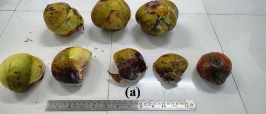
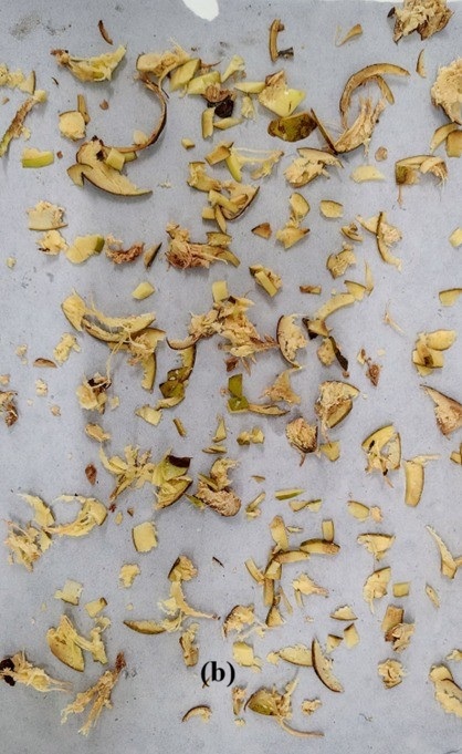
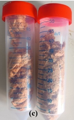
Figure 1. Preparation of plant extract. (a) Raw plant fruit before processing. (b) Separated pericarp part of the fruit. (c) Dried, lyophilized aqueous fruit extract
Phytochemical analysis, UV-Visible, FTIR and GCMS analysis of fruit extract
The extract (EAE) was analysed for the presence of preliminary phytochemicals31 to identify the major classes of secondary metabolites. Further characterization of the extract was carried out by using UV-Visible spectroscopy, Fourier-transform infrared (FTIR) spectroscopy for functional group identification and GC-MS analysis for identification of constituents. The sample for UV-Visible analysis was prepared by dissolving lyophilized extract (1 mg) in 100 mL ethanol and shaken at 200 rpm at room temperature for 2 hr, then filtered to obtain a clear solution. A 1 cm path length quartz cuvette was used in a double beam UV-Vis spectrophotometer (Systronics 2201), with ethanol as the blank. The absorbance spectrum was scanned from 200-800 nm, and measurements were performed in triplicate for reliability. The FTIR analysis was conducted using a Perkin-Elmer Spectrum Two instrument with Universal ATR and Spectrum 10.5.2.636 software. The FTIR spectra of EAE was recorded in the approximately 4000 to 500 cm-1 range. The absorption spectra display characteristic peaks corresponding to various functional groups present in the sample.
Gas Chromatography-Mass Spectrometry (GC-MS) analysis of EAE was conducted using an Agilent MassHunter GC/MS System, operated under MassHunter software for acquisition and data processing. 1 µL aliquot was injected onto an HP 5 capillary column under standard column temperature programming and carrier gas flow conditions. The resulting mass spectra were matched against spectral libraries using the built-in MassHunter. Major compounds are identified on the basis of peak area percentages and retention times under full-scan acquisition mode. The method allowed reliable profiling of volatile and semi-volatile bioactive compounds in the extract.
Determination of total alkaloid content
Total alkaloid content was determined by using the bromocresol green (BCG) spectrophotometric method, as described by Shamsa et al.,32 with atropine as the reference alkaloid.
Preparation of reagents
Bromocresol green solution (BCG) and Phosphate buffer (pH 4.7) solution were prepared as per the Indian Pharmacopoeia. Standard atropine solution was obtained by dissolving 1 mg of pure atropine in 10 ml of distilled water (stock I).
Preparation of standard graph
Accurate measure of aliquots (0.4, 0.6, 0.8, 1 and 1.2 mL) of stock I transfer in a separating funnel. 5 mL of each phosphate buffer and BCG solution were mixed and shake well. A yellow coloured complex was formed. Then extract with 1, 2, 3 and 4 mL of chloroform (in parts 1+2+3+4 = 10 mL) step by step and transfer into a volumetric flask (10 mL). The absorbance was measured at 470 nm in UV spectrophotometer (SHIMADZU UV-1800) against the blank. The blank sample was prepared in the same manner without Atropine.
Preparation of sample
10 gm of extract (EAE) was dissolve in 2N HCL to make the pH upto 2 and then filtered. 1 mL of solution was transferred to a separating funnel and then extracted with 10 mL chloroform (10X3 times). Sodium hydroxide (0.1N) was used to make the pH mixture to neutral. Then 5 mL each of BCG solution and phosphate buffer were added. The mixture was vortexed and the complex formed was extracted with 1, 2, 3 and 4 mL chloroform by gentle shaking. The extracts were collected in a 10 mL volumetric flask and make up the volume upto 10 mL with chloroform. The absorbance was measured at 470 nm.
Determination of Total Phenolic Content (TPC)
The TPC of the fruit extract was determined using the Folin–Ciocalteu colorimetric method as per Singleton et al.33
Preparation of reagents
Folin-Ciocalteu reagent was diluted with distilled water in the ratio of 1:10 (v/v). 7.5% w/v of Sodium carbonate solution (Na2CO3) was prepared. As a standard gallic acid (1 mg/10 ml of distilled water) was used.
Preparation of standard graph
Aliquots of 0.2 ml, 0.4 ml, 0.6 ml, 0.8 ml and 1 ml were transferred into separate test tubes, then the volume was adjusted to 1 ml with distilled water. To each tube, 5 ml of diluted Folin-Ciocalteu reagent was added and mixed thoroughly. After a period of 5 minutes, Na2CO3 solution (7.5% w/v; 4 ml) was mixed to every test tube & vortexed. The reaction mixtures were incubated approximately at 25 °C in darkness for half an hour for colour complex development. The absorbance was measured at 765 nm using a UV spectrophotometer (SHIMADZU UV-1800) against the blank. The blank sample was prepared in the same manner without gallic acid.
Preparation of sample
About 0.2 mL of the EAE (1 mg/mL) was dissolved in Folin–Ciocalteu reagent (1 mL). The mixture was kept to incubate for 5 minute at 25 °C. After that 0.8 mL of Na2CO3 was added. The incubation was carried out for 30 minutes at 25 °C in darkness to allow colour development. The absorbance of the sample solution was measured at 765 nm using visible spectrophotometer against the reagent blank. The total phenolic content was calculated from the calibration curve of gallic acid.
Determination of Total Flavonoid Content (TFC)
TFC was determined using the colorimetric assay as per Chang.34 Quercetin served as the standard compound.
Preparation of reagents
Sodium nitrite (NaNO₂) solution (5% w/v) was prepared. 10% w/v Aluminium chloride solution (upto 100 ml) and Sodium hydroxide (NaOH) solution (1 M) were prepared.
Preparation of standard graph
For the standard solution, quercetin solution (1 mg/ml; methanol) is used and serial dilutions are made to achieve concentrations ranging from 10-100 µg/mL. After that, a quick spin was provided for proper mixing, and a beautiful yellow color obtained.
Preparation of sample
For the reaction mixture, 0.5 mL of the extract or standard solution is pipetted into a test tube, followed by the addition of 2 mL of distilled water and 0.15 mL of the 5% sodium nitrite solution. The mixture is vortexed and allowed to stand for 5 minutes, after which 0.15 mL of the 10% aluminum chloride solution is added and mixed, followed by another 5 minute incubation allow for color reaction. Then, 1 mL of the 1 M sodium hydroxide solution is added, and the mixture is diluted with 0.2 mL of distilled water and mixed thoroughly. The reaction mixture is incubated at 25 °C for half an hour to allow the development of the yellow-colored flavonoid complex. The absorbance of the reaction mixture is measured at 415 nm using a spectrophotometer. A blank control is prepared by replacing the plant extract or standard solution with methanol. The TFC is determined by constructing a calibration curve of absorbance versus quercetin concentration.
Microbial strain and condition of Growth
E. coli (MTCC 242) was used in the present study and maintained in Luria-Bertani (LB) broth media (Himedia). The bacterial culture was first revived by streaking the stock preserved at -80 °C in glycerol onto Luria-Bertani agar (LBA) plates. A well-isolated colony from the plate was then transferred into LBB medium and incubated at 37 °C for 24 hrs. The resulting culture was adjusted to obtain a bacterial suspension of approximately 106 CFU/mL, which was used for all subsequent experiments.
Determination of Minimum Inhibitory Concentration (MIC)
MIC of EAE and gentamicin against E. coli MTCC 242 were determined by using standard broth microdilution assay as outlined by The Clinical & Laboratory Standards Institute (CLSI). Experiments were performed in triplicate.35,36
Bacterial growth curve identification and analysis
To evaluate the antibacterial activity of EAE and gentamicin, the growth kinetics of E. coli MTCC 242 were monitored in the presence and absence of sub-MIC doses of each treatment compared to an untreated control. Briefly, 10 μl of a standardized inoculum (106 CFU/ml) was added to 96-well microtitre plates containing 250 μl of Luria-Bertani Broth (LBB) supplemented with the sub-MIC concentrations. The incubation of the cultures were done at 37 °C, with bacterial growth quantified by recording the optical density (OD) at 590 nm at 6 hrs intervals upto 48 hrs duration.36,37
Estimation of biofilm forming capability of E. coli
Prior to the antibiofilm assay, the biofilm-forming capacity of E. coli MTCC 242 was validated by culturing the organism in Luria-Bertani Broth (LBB). The temperature was maintained at 37 °C for 48 hours. After incubation, the planktonic cells were carefully discarded. The adherent biofilm was then gently rinsed with sterile phosphate-buffered saline (three times; PBS, pH 7.0) to remove any non-attached cells. The remaining biofilm layer was subsequently stained with 0.1% (v/v) safranin for 10 minutes. After removing extra stain with PBS and drying the samples overnight at 37 °C, the safranin bound to the adherent cells was solubilized in 30% (v/v) glacial acetic acid.36,38 The resulting absorbance at 492 nm was measured with the help of PerkinElmer UV-Vis spectrophotometer, with all experiments conducted in triplicate to ensure reproducibility.
Determination of anti-biofilm activity of EAE and gentamicin
The interference of biofilm formation by EAE and gentamicin was evaluated by treating E. coli MTCC 242 with sub-MIC doses (2.5 μg/ml) of each agent and incubating the suspensions at 37 °C for 48 hours. After that, both treated and untreated control tubes were processed according to the previously described biofilm assay, which involved washing, staining with safranin, and measuring the absorbance (OD) at 492 nm. The resulting data were then used to calculate biofilm inhibition (%) in the treated samples from control, providing a quantitative measure of each compound’s antibiofilm efficacy.36-38
Determination of E. coli biofilm total protein after treatment with EAE
To assess the biofilm population density of E. coli MTCC 242 on test tube surfaces, the extractable protein concentration was quantified (directly proportional to microbial density) following treatment with EAE (sub-MIC doses). After incubating the bacteria at 37 °C for 48 hours in the presence or absence of the extract, free floating cells were discarded and remaining attached biofilm cells were gently washed with PBS. The adherent cells were then boiled for 30 minutes in N/2 NaOH to facilitate protein extraction, followed by centrifugation at 10,000 rpm for 5 minutes. Finally, the clear supernatant was collected, and using the Lowry method the total protein concentration was determined to compare the density of treated biofilms against the untreated controls.36,39,40
Microscopic observation of EAE treated E. coli biofilm
To examine the impact of EAE and gentamicin on formation of biofilm over glass surfaces, E. coli was inoculated into 35 × 10 mm Petri dishes with sterile glass coverslips. For incubation temperature maintained (37 °C) for 48 hours with and without of sub-MIC doses. Following incubation, the coverslips were recovered and PBS (sterile) was used for washing and then stained with acridine orange (4 μg/ml) for 15 minutes in the darkness to label the cells. Once dried, the live cells within the adherent biofilm were visualized using a Leica DM 4000B fluorescence microscope,36,41-44 with representative images captured from 20 different fields to ensure a comprehensive assessment of the treatment’s effect.
Measurement of E. coli exopolysaccharide (EPS) after treatment with EAE
To quantify the extracellular polymeric substances (EPS), the biofilm formed on the glass surface was first extracted into water (sterile) and centrifuged at 3,500 g for 20 minutes at 4 °C. To quantify the extracellular polymeric substances (EPS), the formed biofilm on glass surface was first extracted into sterile water and spinned at 3,500 g for 20 minutes at 4 °C. The resulting cell suspension was then suspended with 10 mM/L EDTA with vortex for 15 minutes, and re-centrifuged to isolate cell-bound EPS, with this supernatant being pooled with the initial fraction. To precipitate the EPS, 2.2 volumes of ethanol (absolute, chilled) were added to the pooled supernatant followed by a 1-hour incubation at -20 °C and a final centrifugation step (3,500 g 20 minutes, 4 °C). The recovered EPS pellet was subsequently dissolved in sterile water, and the total carbohydrate content was determined using the phenol–sulfuric acid method.36,45,46
Observation of E. coli motility
To evaluate the impact of EAE and gentamicin on bacterial movement, the swarming motility of E. coli MTCC 242 was investigated using 35 × 10 mm polystyrene plates containing 8 g/l nutrient agar supplemented with 5.0 g/l glucose. An aliquot of 2 μl (treated or untreated) overnight cultures was point-inoculated into the centre of the plates, dry for 20 minutes under laminar blow and subsequently incubated at 37 °C for 48 hours. The motility was then quantified from the diameter of the bacterial migration from the original point of inoculation.47-49
Analysis of statistical parameters
To ensure statistical confidence, experiments were performed in triplicate, with results expressed as the mean ± standard deviation. Statistical significance was evaluated using one-way ANOVA and categorized by P-values of <0.01 (*), P-value <0.001 (**) and P-value <0.0001 (***) while the relationship between different datasets was quantified using the Pearson correlation coefficient (r). All statistical analyses and graphical representations were generated using GraphPad Prism 6.0 software.
Phytochemical analysis
The preliminary phytochemical screening of the EAE reveals the presence of several secondary phytoconstituents like glycosides, flavonoids, tannins, etc. Table 1 outlines the class of metabolites present in the extract.
Table (1): Phytochemical screening of secondary metabolites of fruit extract
| Secondary metabolite | Chemical test | Result | Secondary | Chemical test metabolite | Result |
|---|---|---|---|---|---|
| Alkaloids | Dragendroff’s test and Wagner’s test | + | Phenolic compounds | Ferric chloride test and Folin coicalteu reagent | ++ |
| Flavonoids | Shinoda’s test | ++ | Tannins | Ferric chloride | ++ |
| Alkali (sodium hydroxide) test | ++ | Cardiac glycosides | Legal test | ++ | |
| Lead acetate test | ++ | Steroidal moiety | Libberman’s burchards test | + | |
| Sodium hydroxide test | ++ | Saponins | Foam test | ++ |
+ indicates presence of metabolites, ++ indicates in presence of metabolites with intense colour/ppt
UV-Visible and FTIR Spectroscopy
The UV visible absorption spectrum of the EAE (Figure 2) reveals a prominent absorbance maximum between 220 and 230 nm, reaching values above 2 a.u. This intense peak is characteristic of π-π* electronic transitions strongly suggest the presence of aromatic systems.
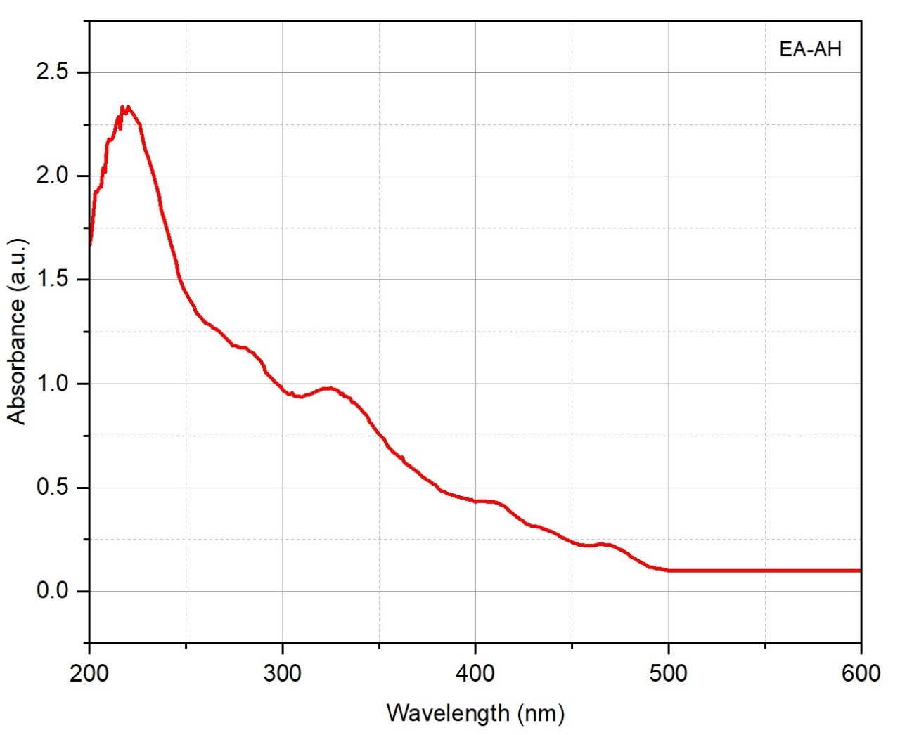
Figure 2. UV–Visible absorption spectrum of Dillenia indica fruit extract
The FTIR spectroscopic analysis (Figure 3) EAE also support the occurrence of polyphenol, flavonoids a carbohydrate related compounds. Major peaks were observed around 2920 cm-1 (C-H stretching of aliphatic chains), 1735 cm-1 (C=O stretching of esters or carboxylic acids), and 1620 cm-1 (C=C stretching of aromatic rings). The additional bands at 1410, 1260 and 1050 cm-1 revealed the presence of O-H bending, C-O stretching and also glycosidic linkage.
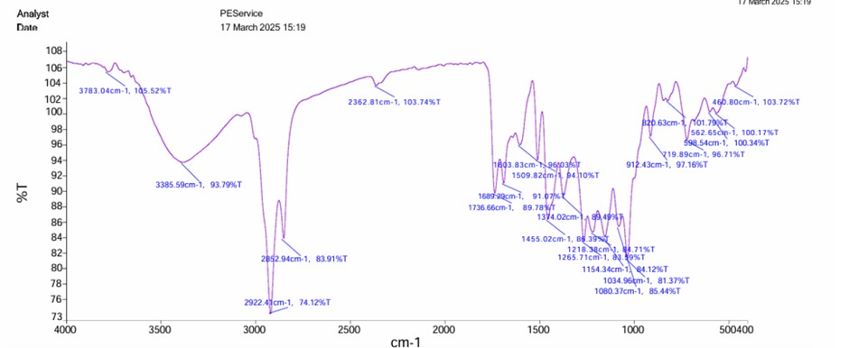
Figure 3. FTIR spectrum of Dillenia indica fruit extract
GC-MS analysis
A total of 33 compounds were reported ranging from small polar organics to more complex esters, heterocycles, and possible artifacts. The compounds are identified via mass spectral library matching (NIST20.L), with similarity scores used to confirm confidence in assignment. The Table 2 outlines the identified compounds by GC-MS.
Table (2): GC-MS identified compounds of Dillenia indica fruit extract
No. |
Compounds |
Mol. formula |
RT |
CAS |
Score |
|---|---|---|---|---|---|
1 |
Butyl isocyanatoacetate |
C7H11NO3 |
3.187 |
17046-22-9 |
78 |
2 |
Hydroxy urea |
CH4N2O2 |
3.511 |
127-07-1 |
73.42 |
3 |
Urea |
CH4N2O |
3.905 |
57-13-6 |
83.73 |
4 |
Silacyclobutane |
C3H8Si |
4.664 |
1000434-34-1 |
74.43 |
5 |
Boronic acid, ethyl- |
C2H7BO2 |
5.711 |
443-63-0 |
70.57 |
6 |
Caprolactone oxime,(NB)-O-[(diethylboryloxy)(ethyl) |
C12H25B2NO2 |
5.933 |
1000161-37-6 |
58.25 |
7 |
Furfural |
C5H4O2 |
6.069 |
98-01-1 |
74.76 |
8 |
Thiophene-3-ol, tetrahydro-,1,1-dioxide |
C4H8O3S |
7.116 |
13031-76-0 |
58.04 |
9 |
Methyl(1H-1,2,3,4-ylmethyl)amine |
C3H7N5 |
7.581 |
1000444-24-2 |
63.04 |
10 |
Octane, 1-azido- |
C8H17N3 |
7.652 |
7438-05-3 |
64.21 |
11 |
5-, alpha.- Aminoethyltetrazole |
C3H7N5 |
7.728 |
1000227-36-7 |
69.12 |
12 |
Cyclohexanol,2-isocyano-,trans- |
C7H11NO |
7.963 |
83152-97-0 |
58.49 |
13 |
Methyl 5-methyl-1H-Carboxylate |
C5H7N3O2 |
8.234 |
1000436-60-0 |
73.46 |
14 |
4H-Pyran-4-one, 2,3-dihydro-3,5- dihydroxy-6-methyl- |
C6H8O4 |
8.540 |
28564-83-2 |
57.75 |
15 |
Pyrazine, methyl-, 1,4-dioxide |
C5H6N2O2 |
8.640 |
32046-26-7 |
49.65 |
16 |
10-Azido-1-decanethiol |
C10H21N3S |
9.128 |
1152113-44-4 |
65.44 |
17 |
Diazene, bis(3-methylcyclohexyl)-1,2- Dioxide |
C14H26N2O2 |
9.357 |
56052-56-3 |
63.71 |
18 |
(4-Isopropyl-1,2,3-triazol-1-yl)acetic acid |
C7H14N3O2 |
9.475 |
1000434-83-8 |
68.71 |
19 |
Bicyclo[3.3.0] octane, 5-dioxa-1-ethyl- |
C6H14BNO2 |
9.569 |
4542-54-5 |
67.91 |
20 |
2,2,5-trimethyl-3,4- dihydro-2h-pyrrole1- oxide |
C7H13NO |
10.252 |
4567-18-4 |
58.36 |
21 |
Hexane,1,6- diisocyanato |
C8H12N2O2 |
10.699 |
822-06-0 |
70.53 |
22 |
1,2,4,5-Tetrazine-3,6-diamine, 1,4-dioxide |
C2H4N6O2 |
11.163 |
153757-93-8 |
70.40 |
23 |
Thiophene-3-ol, tetrahydro-, 1,1-dioxide |
C4H8O3S |
11.163 |
13031-76-0 |
60.80 |
24 |
1,3,2-Dioxaborolane, 2-ethyl-4-(3- oxiranylpropyl) |
C9H17BO3 |
11.699 |
74810-66-5 |
62.69 |
25 |
2,6-Dimethylcyclohexyl methyl phosphonofluoridate |
C9H18FO2P |
12.034 |
1005276-38-9 |
72.27 |
26 |
Nona-2,3-dienoic acid, ethyl ester |
C11H18O2 |
12.993 |
1000187-19-2 |
70.61 |
27 |
1,3,2-Oxazaborolane, 2-butyl- |
C6H14BNO |
13.704 |
31748-10-4 |
63.93 |
28 |
Borinic acid, diethyl-, 5-hexynyl ester |
C10H19BO |
15.839 |
62338-11-8 |
59.65 |
29 |
4-Heptanone, oxime |
C7H15NO |
17.510 |
1188-63-2 |
59.91 |
30 |
4-(1H-1,2,3,4Tetrazol-5- yl) piperidine |
C6H11N5 |
17.228 |
112626-97-8 |
60.90 |
31 |
1,2,5-Oxadiazol-3- carboxamide, 4,4′- azobis-, 2,2′-dioxide |
C6H4N8O6 |
26.868 |
1000294-17-4 |
53.34 |
32 |
Arsenous acid, tris(trimethylsilyl) ester |
C9H27AsO3Si3 |
31.086 |
55429-29-3 |
45.67 |
33 |
Tris(tert- butyl dimethyl lsilyloxy)arsane |
C18H45AsO3Si3 |
40.609 |
100366-57-5 |
63.09 |
Quantitative estimation of alkaloid, phenolic and flavonoid contents
In our current study though alkaloid is qualitatively identified in fruit extract but the quantitative analysis reveals that alkaloid is not a major constituent present in fruit. Phenolic compounds are reported as the major bioactive constituents in the fruit. Flavonoids in the fruit have been reported in moderate amounts and are associated with antimicrobial and anti-inflammatory activities. The quantitative estimation of total alkaloid, total phenolic, and total flavonoid contents are tabulated in Table 3.
Table (3): Quantitative estimation of phytoconstituents
Parameter |
Absorbance (Mean± SEM) |
Content (Mean± SEM) |
Equivalent standard |
|---|---|---|---|
Total alkaloidal content |
0.40 ± 0.01 |
24.50 ± 0.78 µg AE/mL |
Atropine |
Total phenolic content |
0.35 ± 0.01 |
110 ± 3.2 µg GAE/mL |
Gallic acid |
Total Flavonoid content |
0.45 ± 0.02 |
11.28 ± 0.45 µg QE/mL |
Quercetin |
Values are expressed as Mean ± SEM (n = 3). GAE = Gallic acid equivalents; QE = Quercetin equivalents; AE = Atropine equivalents
Antibacterial effect of extract on planktonic form of E. coli
EAE under experimental condition has executed antibacterial effect against E. coli where maximum effect was observed at 540 µg/m concentration.27,28 Antibacterial efficacy of EAE on planktonic bacterial growth against E. coli was 68.25% and 75.12% with 6.23 mm and 6.95 mm (Figure 4) zone inhibition diameter at the concentrations of 270 µg/ml and 540 µg/ml, respectively. Gentamicin as the standard antibiotic exhibited maximum 98.87% inhibition (Figure 5) with 7.9 mm zone inhibition diameter at 1.25 µg/ml concentration. This indicates that EAE has potentiality to induce significant antibacterial potentiality.
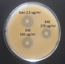
Figure 4. Bacterial zone of inhibition over agar plates in presence of EAE and gentamicin
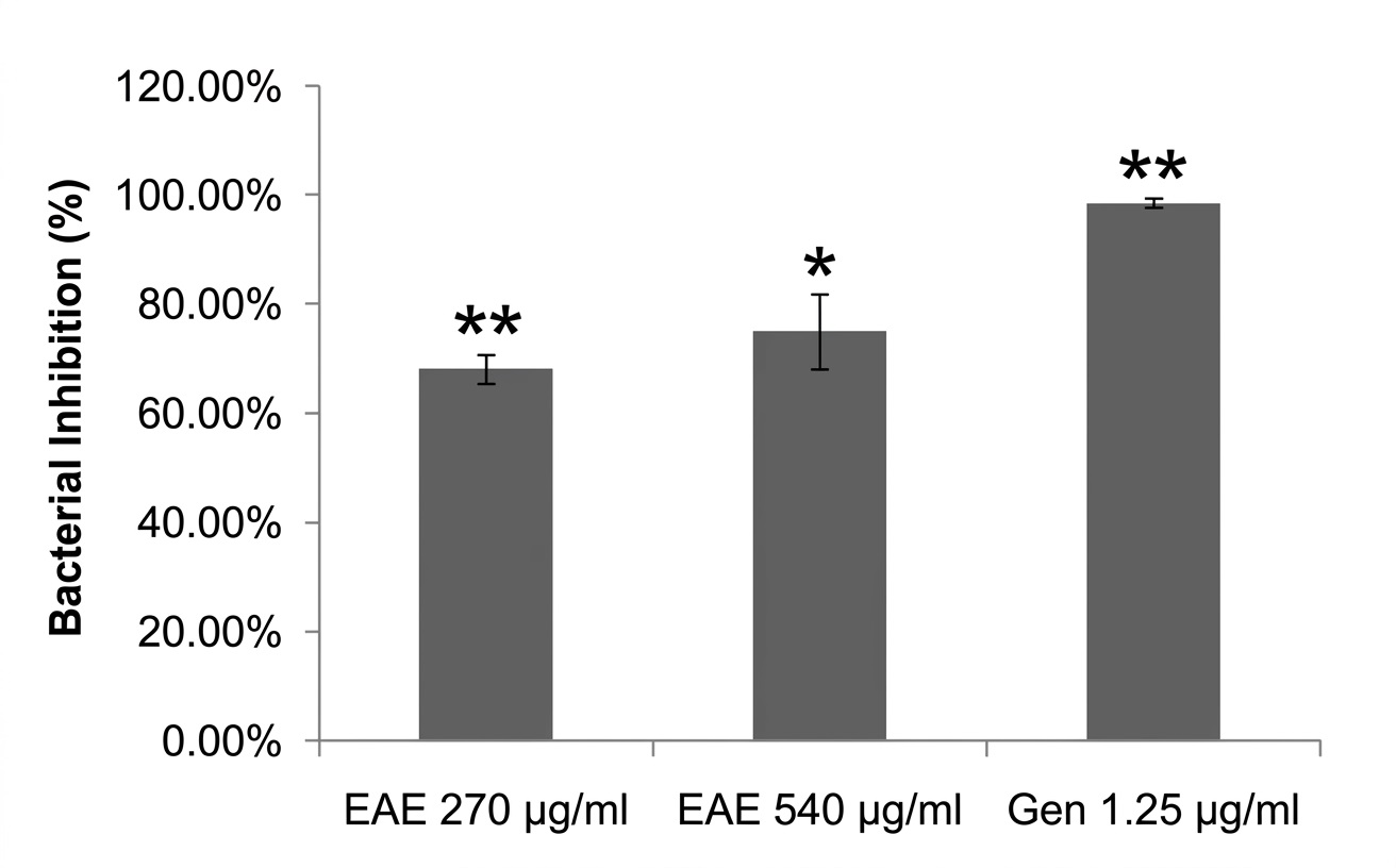
Figure 5. Inhibition (%) of planktonic bacteria with respect to untreated control
Anti-biofilm effect of EAE on E. coli biofilm
Anti-biofilm efficacy studies of EAE showed a substantial biofilm attenuation (66.27%) of E. coli at 270 µg/ml concentration compared to gentamicin, which, as the standard antibiotic showed 90.93% attenuation at a sub-inhibitory dose of 1.25 µg/ml (Figure 6) manifesting the significant bacterial biofilm inhibition capability of EAE. In addition, it was also observed that EAE significantly reduced E. coli dispersion at 270 µg/ml concentration from preformed biofilm (Figure 6). Further, in fluorescent microscopic study it was noticed that bacterial aggregation was also greatly inhibited by EAE corroborating its biofilm inhibition against E. coli (Figure 7). It was also found that at certain sub-inhibitory concentration, these EAE start dispersing planktonic cells from biofilm to dislodge the network architecture. In expressing result of antibacterial and anti-biofilm R2 value were shown to express linearity of data plotted in the curve and significance of data in the series of concentrations that were studied. The effects of EAE on biofilm network were also observed under fluorescent microscope with respect to untreated control and gentamicin as reference antibiotic to inhibit formation of biofilm cluster.44-47
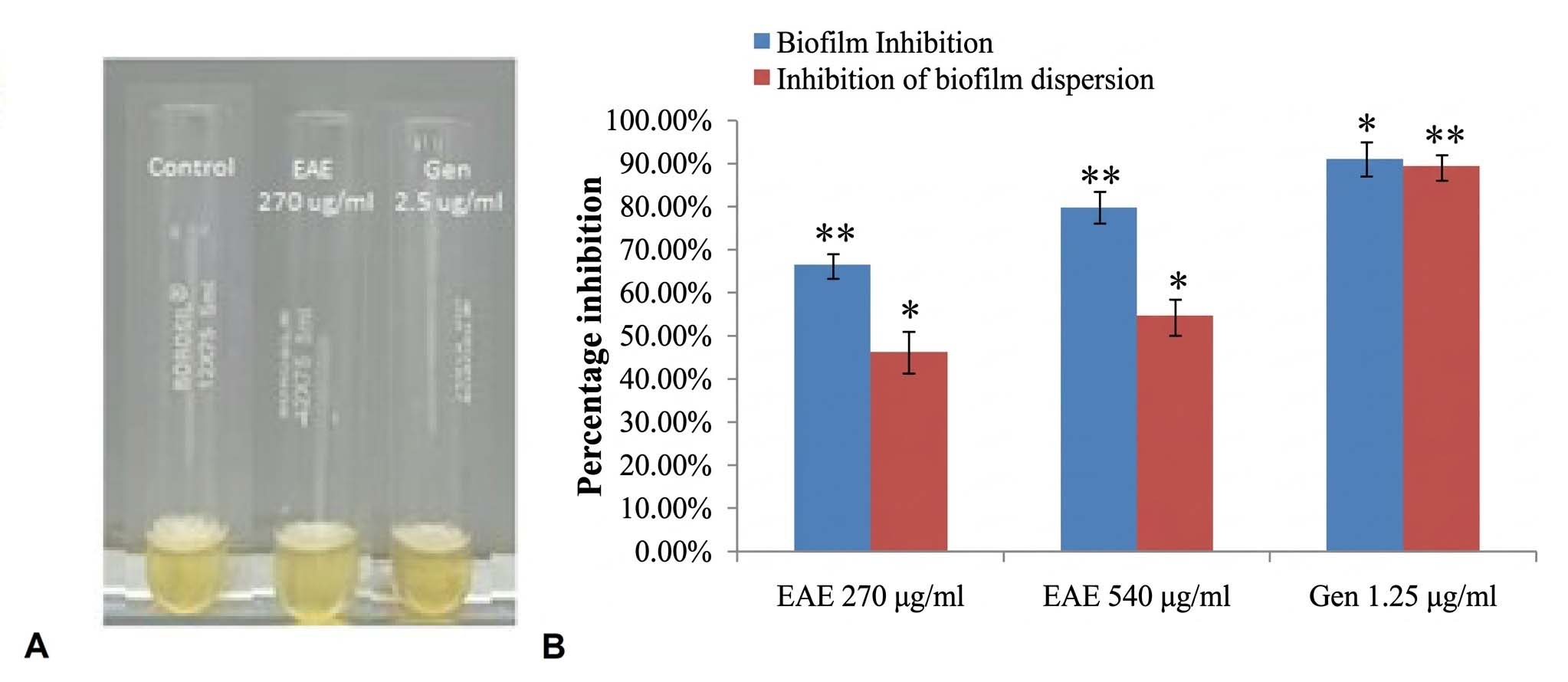
Figure 6. Bacterial biofilm inhibition (A). Graphical representation of biofilm inhibition and inhibition of biofilm dispersion (B)
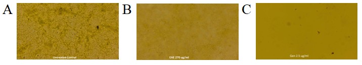
Figure 7. Fluorescent microscopic images of E. coli biofilm inhibition. (A) With respect to untreated control. (B) After treatment with EAE. (C) After treatment with gentamicin
Effect of EAE on bacterial motility
In motility assay, EAE exerted significant motility reduction (Figure 8) of bacterial cells compared to the reference standard. It was also observed that bacterial colonization (primarily required for biofilm formation) was notably disrupted indicating the capability of biofilm inhibition by EAE at sub-inhibitory concentrations. In the study of bacterial motility, we have observed that 270 µg/ml EAE treated cells showed marked reduction in sliding movement compared to the negative control (Figure 8), however, sub-MIC dose of gentamicin (1.25 µg/ml) also showed very significant attenuation of bacterial motility.
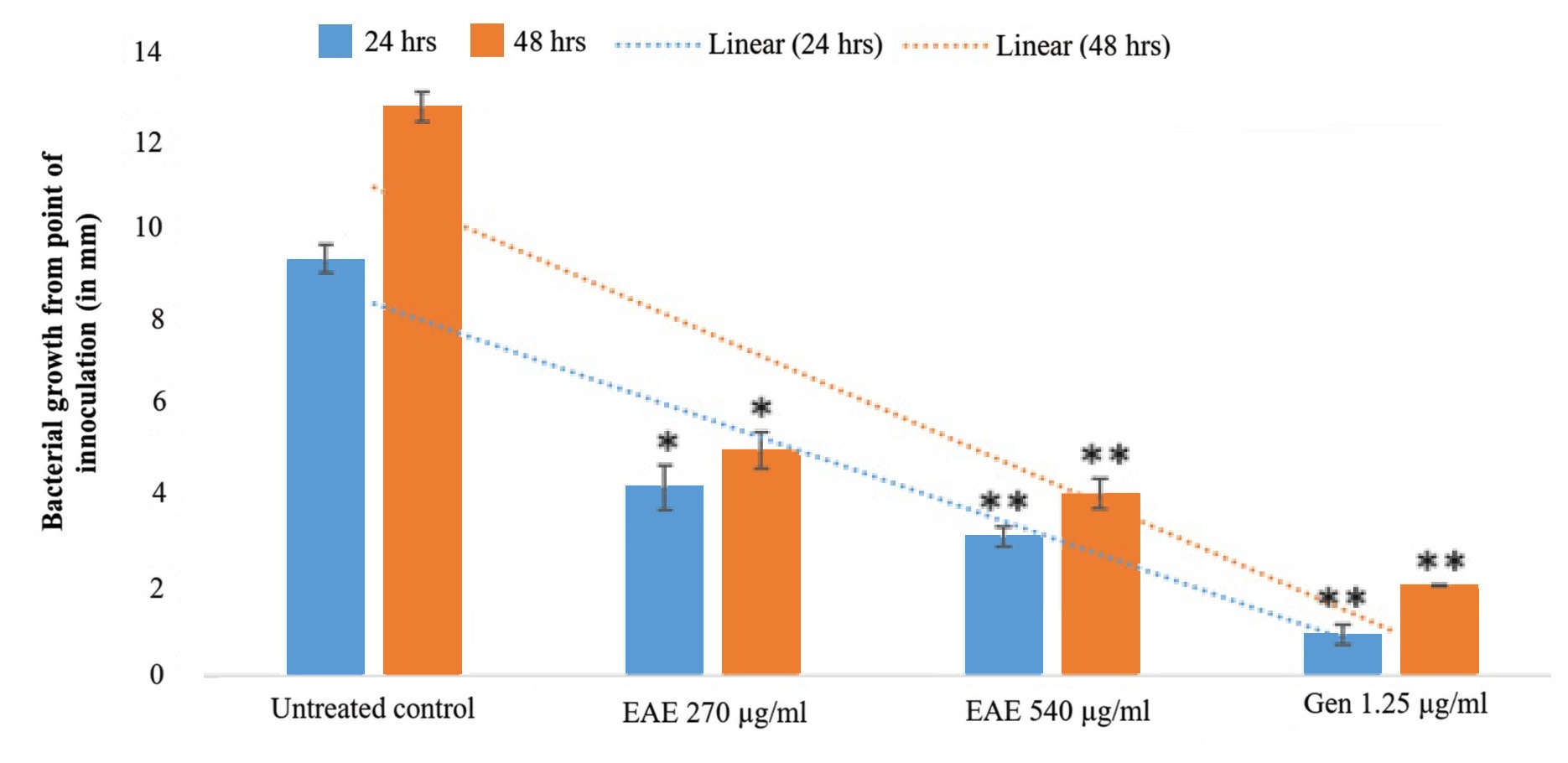
Figure 8. Graphical representation of growth of bacteria from their point of inoculation
Estimation of bacterial EPS
In the present work we have quantified the amount of EPS in all experimental sets as per the scheme of the work. We have observed a significant correlation (r = 0.899754) in the measured quantity of EPS with anti-biofilm assays. The results indicated that samples treated with EAE at 270 µg/mL exhibited a significantly lower amount of EPS, whereas the untreated control samples showed the highest level of EPS production. (Figure 9). Furthermore, after the combined action of sub-MIC doses of EAE and gentamicin, EPS quantities were reduced significantly which exhibited 73.69% inhibition (Figure 9). The results were expressed as percentage of inhibition relative to the untreated control. Furthermore, the ratio of biofilm protein and EPS quantity reveals that with treatment reduction rate of protein was higher than EPS but maintains a steady state. All these results validated that EAE has potent anti-biofilm activity at sub-MIC dose.
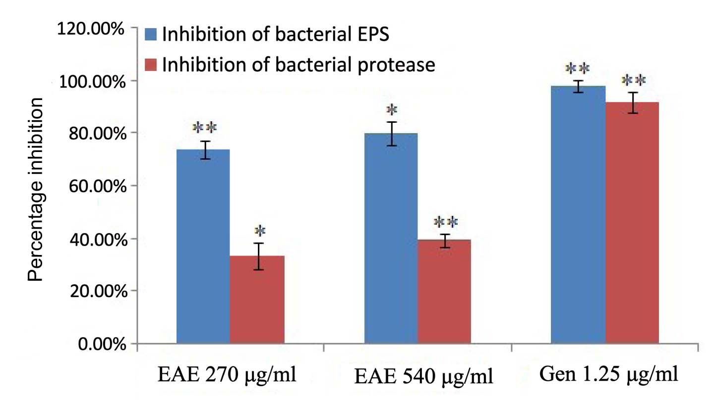
Figure 9. Graphical representation of inhibition (%) of bacterial EPS and proteases
The presence of secondary metabolites is the key factor for any therapeutic activity. In the current study, qualitative analysis of aqueous extract of the elephant apple showed the presence of phenolic compounds, flavonoids, saponins, alkaloids, etc. which support its therapeutic activity. In UV analysis, the electronic transition strongly suggests a high abundance of phenolic compounds, including ellagic acid and related polyphenols. Such transitions are typically confirmed the presence of phenolic acids and certain flavonoid aglycones as already confirmed by Alam et al.43 A broad shoulder observed in the 300-350 nm range (with absorbance of ~0.8-1.0 a.u.) likely reflects the presence of conjugated systems including flavonols and hydroxycinnamic acids, such as quercetin derivatives and other flavonoids previously identified in the plant.43,50-52 The continuous decline in absorbance across the visible region with the spectrum approaching baseline near 500 nm indicates a relative paucity of strongly colored plant pigments, such as chlorophylls or carotenoids. Collectively, the spectral features are consistent with the established phytochemistry of the plant, which is rich in polyphenols (including ellagic acid, gallic acid), flavonoids50-52 (quercetin, kaempferol), and to a lesser extent, terpenoid and glycosidic compounds. The absence of significant absorption in the visible region further supports the predominance of colorless or pale-colored phenolic constituents over other chromophores. These observations support and complement published reports on the diverse polyphenolic and flavonoid content of the plant,28 providing a spectral fingerprint that can be used as a qualitative indicator of these bioactive phytochemicals.
In the current study, FTIR peaks strongly suggest the presence of polyphenolic compounds, such as flavonoids and tannins. Such metabolites are known for their antioxidant and antimicrobial properties.53-55 The prominent peak near 2920 cm-1 is attributed to C-H stretching vibrations of aliphatic chains, indicative of alkanes commonly found in triterpenoids or fatty acid components of the plant extract.56 A distinct absorption band at 1735 cm-1 corresponds to C=O stretching vibrations characteristic of esters or carboxylic acids, functional groups typically present in triterpenoids and flavonoid glycosides reported in this plant. An absorption band around 1620 cm-1 indicates C=C stretching vibrations within aromatic rings, consistent with flavonoids and phenolic acids, supporting their abundance in the extract. The peak near 1410 cm-1 may be due to O-H bending or C-H bending vibrations, common in flavonoid structures and carboxylic acids, reinforcing the presence of polyphenolic compounds. The band near 1260 cm-1 corresponds to C-O stretching, associated with esters and secondary alcohols, likely corresponding to flavonoid or triterpenoid derivatives. A strong absorption peak around 1050 cm-1 corresponds to C-O-C stretching vibrations, suggestive of glycosidic bonds commonly found in polysaccharides and flavonoid glycosides. Overall, the FTIR spectral profile confirms the presence of polyphenols, flavonoids, glycosides, triterpenoids, and carbohydrates in the fruit extract. These findings correlate well with previously reported secondary metabolites of this plant and provide a chemical basis for its observed biological activities. Since compounds such as flavonoids, phenolic acids, and triterpenoids are known for their antimicrobial and anti-biofilm properties,57,58 the identification of these functional groups supports the plant extract’s anti-biofilm activity.59
In the current study GCMS peaks with high-intensity were associated with common phytochemicals such as urea, furfural, and nonanoic acid derivatives. Several research claims the activity of urea, nonanoic acid derivatives as antimicrobial60-62 and fatty acids as anti-biofilm and anti-virulence agents.63 There are several other less conventional plant constituents are present for e.g. silacyclobutane and boronic acid derivatives. The multiple times presence of urea could indicate that urea (either as an endogenous metabolite or as an adulterant) may have diagnostic value for metabolic or environmental stress in plants and their products.41 Further, the presence of furfural may associate to flavor and aroma profile of the fruit. Presence of sulfone derivatives could consider as environmental contaminants and a prime factor for further research. Detection of butyl isocyanatoacetate may be attributable to agricultural practices or potentially indicate an unusual or previously unreported biosynthetic pathway within the plant. The occurrence of organosilicon compounds points to possible interactions between the plant and the surrounding soil or other environmental sources. Additionally, the recurrent detection of arsenic compounds highlights biogeochemical processes unique to the sampling location and may suggest novel arsenic detoxification mechanisms in the plant.
The quantitative analysis revealed the presence of alkaloids, phenolics, and flavonoids in the aqueous fruit extract. The presence of alkaloids in leaf extract has already been identified by the Biswas et al. in the year 2015.64 Studies also reported the presence of berberine as one of the alkaloid in the plant, which is responsible for antimicrobial activity against several bacterial infections.65,66 Thus, the presence of alkaloids may also contribute to the antibacterial and antibiofilm activities, in addition to the phenolic and flavonoid compounds present in the extract. The phenolic content obtained in the present study is consistent with earlier reports indicating that Dillenia indica fruit is rich in polyphenolic compounds. Previous studies have demonstrated that aqueous and hydroalcoholic extracts of the fruit contain significant levels of phenolic compounds responsible for antioxidant and antimicrobial activities. Biswas et al.64 also revealed the presence of flavonoids like quercetin and kaempherol in the plant. Furthermore, previous studies also express the presence of higher phenolic content in the methanol fruit extract (approximately 34% of phenolic content expressed as tannic acid equivalent), whereas aqueous extract contains only 1.4%.18,67 The flavonoid content observed in the present study also aligns with previously reported values for aqueous fruit extract, supporting the presence of bioactive flavonoids65 contributing to its pharmacological activities. The variations observed in alkaloid, phenolic, and flavonoid contents as compared to previous studies may be due to the geographical source, collection procedure, and extraction parameters adopted etc.
In the present work bacterial inhibition by EAE was compared with standard antibiotic gentamicin. Gentamicin enters gram-negative bacteria through oxygen-dependent active transport system leading to miscoding of protein and cell death. Gentamicin is very effective antibiotic against E. coli as indicated by its bacterial growth inhibition.38,43 In comparison to that EAE has shown moderate to significant antibacterial effect which acts as baseline data for anti-biofilm study. Observation of the present study suggests that EAE at sub-inhibitory concentrations executed moderate to significant bacterial biofilm attenuation with respect to gentamicin. Such anti-biofilm effects were observed in glass tubes as well as under microscopes. Under magnification, it was observed that EAE has significantly attenuated bacterial aggregation to form biofilm clusters.43-46 These observations suggest that EAE has potentiality to modulate bacterial biofilm formation. Furthermore, it was also observed that EAE at sub-inhibitory concentrations disperse planktonic bacteria from their aggregated state. R2 value of biofilm formation and dispersion data also supports such observations. Bacteria in natural eco-system perform movement from a specific point for collection of nutrient and as part of community behavior.43-46 Bacterial motility is a quorum sensing behavior through which bacteria communicate among themselves and collect food from the environment.38,43-46 It was also observed that under the influence of EAE bacterial motility was also modulated. Thus, attenuation of bacterial motility by sub-inhibitory concentrations of EAE supports attenuation of bacterial aggregation to form biofilm. In a nutshell it can be discussed that EAE has potentiality to modulate bacterial communication system and subsequent attachment of cells to form aggregated biofilm.
The aqueous extract of Elephant apple fruit (EAE) revealed the presence of several functional groups corresponding to phenols, flavonoids, carboxylic acids, esters, triterpenoids, glycosides, and carbohydrates. These chemical groups indicate a rich phytochemical composition consistent with previously reported constituents of the plant. The detection of these functional groups suggests their potential role in the extract’s observed anti-biofilm and antibacterial activities, as supported by the literature. Therefore, the chemical composition obtained from the fruit not only supports the traditional use of Dillenia indica in gastrointestinal disorders but also provides a scientific rationale for its bioactivity, which has been further confirmed through presence of phytochemicals and biological assays in the present study.
ACKNOWLEDGMENTS
The authors acknowledge the Department of MLT, Women`s Polytechnic College, Hapania, Tripura, for providing the facility to carry out the anti-bacterial and anti-biofilm work.
CONFLICT OF INTEREST
The authors declare that there is no conflict of interest.
AUTHORS’ CONTRIBUTION
SC, MCD and AKY conceptualized and designed the study. SC, HS and MCD performed literature review. SC, HS, MCD and AKY performed experiments, data acquisition, data analysis and statistical analysis. SC, HS and MCD wrote, reviewed and revised the manuscript. All authors read and approved the final manuscript for publication.
FUNDING
None.
DATA AVAILABILITY
All datasets generated or analyzed during this study are included in the manuscript.
ETHICS STATEMENT
Not applicable.
- Bordoloi J, Dihingia A, Kalita J, Manna P. Ethnomedicinal Plants of North-East India as a Potential Target for Drug Discovery Against Type 2 Diabetes Mellitus. In: Patra J, Shukla A, Das G. (eds) Advances in Pharmaceutical Biotechnology. Springer, Singapore. 2020:39-54.
Crossref - Dutta SK, Vanlalhmangaiha, Akoijam RS, Lungmuana, Boopathi T, Saha S. Bioactivity and traditional uses of 26 underutilized ethnomedicinal fruit species of North-East Himalaya, India. J Food Meas Charact. 2018;12(2):2503-2514.
Crossref - Gaibimei P, Devi NM, Raleng A, Pongener A, Mate CJ. Sustainable processing and commercialization of underutilized fruits in North East India. Int J Environ Agric Biotechnol. 2025;10(3):99-110.
Crossref - Nutraceuticals market size, share and trends analysis report. https://www.grandviewresearch.com/industry-analysis/nutraceuticals-market, Accessed 12 February, 2026.
- Fruit extracts Market by source (Citrus fruits, apples, pineapples, grapes, pears, berries), form (Powder, liquid), Application (Beverages, food, nutraceuticals, cosmetics) – Global forecast to 2032. Accessed 12 February, 2026 https://www.meticulousresearch.com/product/fruit-extracts-market-5782
- Das M, Choudhury M, Sarma BP. Dillenia indica L. In Belwal T, Bhatt I, Devkota H (eds.), Himalayan Fruits and Berries: Bioactive Constituents, Uses and Nutraceutical Potential. Academic Press. 2023:111- 120.
Crossref - Mesa NC, Alves IA, Vilela FMP, et al. Fruits as nutraceuticals: A review of the main fruits included in nutraceutical patents. Food Res Int. 2023;170:113013.
Crossref - Bhardwaj Y, Bhuyan B, Pulicherla Y, et al. Ethnomedicinal plants used for gastro-intestinal disorders (GIDs) by the tribal communities of Arunachal Pradesh (Eastern Himalayas), India: A comprehensive review. Ethnobot Res Appl. 2025;30(5):1-39.
- Sharma HK, Chhangte L, Dolui AK. Traditional medicinal plants in Mizoram, India. Fitoterapia. 2001;72(2):146-161.
Crossref - Kumar D, Mallick S, Vedasiromoni JR, Pal BC. Anti-leukemic activity of Dillenia indica L. fruit extract and quantification of betulinic acid by HPLC. Phytomedicine. 2010;17(6):431-435.
Crossref - Fu C, Yang D, Peh WY, Lai S, Feng X, Yang H. Structure and antioxidant activities of proanthocyanidins from elephant apple (Dillenia indica Linn.). J Food Sci. 2015;80(10):C2191-C2199.
Crossref - Talukdar N, Talukdar A. Deka S, Sahariah BJ. Dillenia indica (Outenga) as an anti-diabetic herb found in Assam: A review. Int J Pharm Sci Res. 2012;3(8):2482-2486.
Crossref - Kamboj P, Talukdar NC, Banerjee SK. Therapeutic benefit of Dillenia indica in diabetes and its associated complications. J Diabetes Res. 2019;2019:4632491.
Crossref - Kumar S, Kumar V, Prakash O. Enzyme inhibition and antidiabetic effect of isolated constituents from Dillenia indica. Biomed Res Int. 2013;2013:382063.
Crossref - Rahman SS, Meherunnahar, Reja MdM, et al. Proximate nutrient analysis of elephant apple (Dillenia indica) fruit and its hypoglycemic, and hypolipidemic potentials in alloxan-induced diabetic rats. Food Hum. 2023;1:1355-1361.
Crossref - Khare RK, Prasad AK, Kumar S, Iyer SV, Vaidya SK, Bigoniya P. Flavonoid- and triterpene-rich fraction of Dillenia indica Linn. leaves: Anti-inflammatory and anti-arthritic activity. Indo Am J Pharm Res. 2013;3(6):4653-4659.
- Padhya IP, Choudhary NS, Padhy SK, Dash S. Effect of Dillenia indica leaves against carbon tetrachloride-induced hepatotoxicity. J Pharm Chem. 2008;2(4):24-27
- Abdille MH, Singh RP, Jayaprakasha GK, Jena BS. Antioxidant activity of the extracts from Dillenia indica fruits. Food Chem. 2005;90(4):891-896.
Crossref - Yeshwante SB, Juvekar AR, Nagmoti DM, et al. Anti-inflammatory activity of methanolic extracts of Dillenia indica L. leaves. J Young Pharm. 2009;1(1):63-66.
Crossref - Yeshwante SB, Juvekar AR, Pimprikar RB, et al. Anti-diarrheal activity of methanolic and aqueous extracts of Dillenia indica L. Res J Pharmacol Pharmacodyn. 2009;1(3):140-142.
- Rahman MS, Shams-Ud-Doha KM, Rahman R. Antidiarrhoeal activity of the leaf and fruit extracts of Dillenia indica. Int J Biosci. 2011;1(6):39-46.
- Alam MB, Sarowar M, Hossain MdS, Ekramul H. Evaluation of antimicrobial and toxicity of different fractions of Dillenia indica Linn. bark extract. J Glob Pharm Technol. 2010;2(11): 975-8542.
- Khan MA, Ahamad T, Saquib M, Hussain MK, Khan MF. Unmodified household coffee maker-assisted extraction and purification of anticancer agents from Dillenia indica fruits. Nat Prod Res. 2021;35(6):984-987.
Crossref - de Souza AA, Dias Viegas FP, Gontijo VS, et al. Antinociceptive effect of Dillenia indica (Linn.) mediated by opioid and cannabinoid systems: pharmacological and chemical studies. Chem Biodivers. 2024;21(3):e202301508.
Crossref - Reddy KH, Reddy KBN, Sharma PVGK, Reddy OVS. In vitro Studies on Antimicrobial and Antioxidant Activities of Dillenia indica Seed Extract. J Pure Appl Microbiol. 2009;3(2):769-776
- Reddy KH, Govender P, Reddy OVS, Sarma PVGK. Effect of hexane extract of Dillenia indica seed on the activity of secreted aspartyl proteinase of Candida albicans and its kinetic studies. J Med Plants Res. 2012;6(44):5599-5603.
Crossref - Nkop EJ, Zudonu OC. Phytochemical screening and antimicrobial activity of leaves and stem bark of Dillenia indica. Pharm Chem J. 2020;7(1):1-4.
- Jaiswal S, Mansa N, Prasad MP, Jena BS, Negi PS. Antibacterial and antimutagenic activities of Dillenia indica extracts. Food Biosci. 2014;5:47-53.
Crossref - Di Domenico EG, Farulla I, Prignano G, et al. Biofilm is a major virulence determinant in bacterial colonization of chronic skin ulcers independently from the multidrug resistant phenotype. Int J Mol Sci. 2017;18(5):1077.
Crossref - Wang D, Wang L, Liu Q, Zhao Y. Virulence factors in biofilm formation and therapeutic strategies for Staphylococcus aureus: A review. Anim Zoonoses. 2025;1(2):188-202.
Crossref - Khandelwal KR. Practical Pharmacognosy: Techniques and Experiments. 19th Ed. Nirali Prakashan, Pune 2008.
- Shamsa F, Monsef H, Ghamooshi R, Verdian-Rizi M. Spectrophotometric determination of total alkaloids in some Iranian medicinal plants. Thai J Pharm Sci. 2008;32(1):17-20.
Crossref - Singleton VL, Orthofer R, Lamuela-Raventos RM. Analysis of total phenols and other oxidation substrates and antioxidants by means of Folin-Ciocalteu reagent. Methods Enzymol. 1999;299:152-178.
Crossref - Chang CC, Yang MH, Wen HM, Chern JC. Estimation of total flavonoid content in propolis by two complementary colorimetric methods. J Food Drug Anal. 2002;10(3):178-182.
Crossref - Mekonnen KD. Fourier transform infrared spectroscopy as a tool for identifying the unique characteristic bands of lipid in oilseed components: Confirmed via Ethiopian indigenous desert date fruit. Heliyon. 2023;9(4):e14699.
Crossref - Gupta A, Pandey BC, Yaseen M, et al. Exploring anticancer, antioxidant, and antimicrobial potential of Aspergillus flavus, a fungal endophyte isolated from Dillenia indica leaf callus. Heliyon. 2025;11(3):e42142.
Crossref - Bora P, Devi NN. Exploration of the chemical constituents and its antioxidant, antibacterial activities of endophytic fungi isolated from the medicinal plant Dillenia indica. Arch Microbiol. 2023;205(2):67.
Crossref - Patil M, Noonikara-Poyil A, Joshi SD, Patil SA, Patil SA, Bugarin A. New Urea Derivatives as Potential Antimicrobial Agents: Synthesis, Biological Evaluation, and Molecular Docking Studies. Antibiotics 2019;8(4):178.
Crossref - Lee JH, Kim YG, Khadke SK, Lee J. Antibiofilm and antifungal activities of medium-chain fatty acids against Candida albicans via mimicking of the quorum-sensing molecule farnesol. Microb Biotechnol. 2020;14(4):1353-1366.
Crossref - Kumar P, Lee JH, Beyenal H, Lee J. Fatty acids as antibiofilm and antivirulence agents. Trends Microbiol. 2020;28(9):753-768.
Crossref - Haji A, Desalegn K, Hassen H. Selected food items adulteration, their impacts on public health, and detection methods: A review. Food Sci Nutr. 2023;11(12):7534-7545.
Crossref - Luo Y, Song Y. Mechanism of antimicrobial peptides: antimicrobial, anti-inflammatory and antibiofilm activities. Int J Mol Sci. 2021;22(21):11401.
Crossref - Alam MB, Ahmed A, Islam S, et al. Phytochemical characterization of Dillenia indica L. bark by paper spray ionization-mass spectrometry and evaluation of its antioxidant potential against t-BHP-induced oxidative stress in RAW 264.7 cells. Antioxidants. 2020;9(11):1099.
Crossref - Yakandawala N, Gawande PV, LoVetri K, Madhyastha S. Effect of ovotransferrin, protamine sulfate and EDTA combination on biofilm formation by catheter-associated bacteria. J Appl Microbiol. 2007;102(3):722-727.
Crossref - National Committee for Clinical Laboratory Standards. Methods for dilution antimicrobial susceptibility tests for bacteria that grow aerobically. Wayne, PA: National Committee for Clinical Laboratory Standards. 1997.
- Das MC, Sandhu P, Gupta P, et al. Attenuation of Pseudomonas aeruginosa biofilm formation by vitexin: a combinatorial study with azithromycin and gentamicin. Sci Rep. 2016;6:23347.
Crossref - Bauer AW, Kirby WM, Sherris JC, Turck M. Antibiotic susceptibility testing by a standardized single disk method. Am J Clin Pathol. 1966;45(4):493-496.
- Jabra-Rizk MA, Meiller TF, James CE, Shirtliff ME. Effect of farnesol on Staphylococcus aureus biofilm formation and antimicrobial susceptibility. Antimicrob Agents Chemother. 2006;50(4):1463-1469.
Crossref - Lowry OliverH, Rosebrough NiraJ, Farr AL, Randall R. Protein Measurement with the Folin Phenol Reagent. J Biol Chem. 1951;193(1):265-275.
Crossref - Abd El–Kader EM, Shakour ZTAE. Phytochemical and cytotoxicity investigation of Dillenia indica grown in Egypt. World J Pharm Res. 2015;4(10):334-348.
- Aswathy M, Banik K, Parama D, et al. Exploring the cytotoxic effects of extracts and bioactive triterpenoids from Dillenia indica against oral squamous cell carcinoma: A scientific interpretation and validation of indigenous knowledge. ACS Pharmacol Transl Sci. 2021;4(2):834-847.
Crossref - Lawal TO, Raut NA, Patel SR, Mahady GB. Extracts of Anogeissus leiocarpus and Dillenia indica inhibit the growth of MCF-7 breast cancer and COV434 granulosa tumor cells by inducing apoptosis and autophagy. Curr Bioact Compd. 2021;17(10):35-48.
Crossref - Rahman MM, Hunter HN, Prova S, Verma V, Qamar A, Golemi-Kotra D. The Staphylococcus aureus methicillin resistance factor FmtA is a D-amino esterase that acts on teichoic acids. mBio. 2016;7:e02070-15.
Crossref - Collins LV, Kristian SA, Weidenmaier C, et al. Staphylococcus aureus strains lacking D-alanine modifications of teichoic acids are highly susceptible to human neutrophil killing and are virulence attenuated in mice. J Infect Dis. 2002;186(2):214-9.
Crossref - Banin E, Brady KM, Greenberg EP. Chelator-induced dispersal and killing of Pseudomonas aeruginosa cells in a biofilm. Appl Environ Microbiol. 2006;72(3):2064–2069.
Crossref - Schillaci D, Arizza V, Parrinello N, et al. Antimicrobial and antistaphylococcal biofilm activity from the sea urchin Paracentrotus lividus. J Appl Microbiol. 2010;108(1):17-24.
Crossref - Das MC, Paul S, Gupta P, et al. 3-Amino-4-aminoximidofurazan derivatives: small molecules possessing antimicrobial and antibiofilm activity against Staphylococcus aureus and Pseudomonas aeruginosa. J Appl Microbiol. 2016;120(4):842-859.
Crossref - Sabharwal N, Dhall S, Chhibber S, Harjai K. Molecular detection of virulence genes as markers in Pseudomonas aeruginosa isolated from urinary tract infections. Int J Mol Epidemiol Genet. 2014;5(3):125-534.
- Dubois M, Gilles KA, Hamilton JK, Rebers PA, Smith F. Colorimetric method for determination of sugars and related substances. Anal Chem. 1956;28(3):350-356.
Crossref - Harimawan A, Ting YP. Investigation of extracellular polymeric substances (EPS) properties of Pseudomonas aeruginosa and Bacillus subtilis and their role in bacterial adhesion. Colloids Surf B Biointerfaces. 2016;146:459-467.
Crossref - Kumar L, Chhibber S, Harjai K. Zingerone inhibits biofilm formation and improves antibiofilm efficacy of ciprofloxacin against Pseudomonas aeruginosa PAO1. Fitoterapia. 2013;90:73-78.
Crossref - Kaito C, Sekimizu K. Colony spreading in Staphylococcus aureus. J Bacteriol. 2007;189(6):2553-2557.
Crossref - Kessler E, Safrin M, Olson JC, Ohman DE. Secreted LasA of Pseudomonas aeruginosa is a staphylolytic protease. J Biol Chem. 1993;268(10):7503-7508.
- Biswas S, Pandita N. Phytochemical analysis and chromatographic evaluation of alcoholic extract of Dillenia indica Linn. leaves. Int J Pharm Sci Res. 2015;6(7):2799-2812.
Crossref - Nahar L, Habibi E, Khuniad C, et al. Bioactive phytochemicals, pharmacological, and therapeutic potential of Dillenia indica: A comprehensive review of current research. Chin Herb Med. 2025;17(4):628-642.
Crossref - Yang X, Wang Y, Li L, et al. Berberine and its nanoformulations and extracts: Potential strategies and future perspectives against multidrug-resistant bacterial infections. Front Microbiol. 2025;16:1643409.
Crossref - Singh AK, Saha S. Chemistry, Therapeutic Attributes, and Biological Activities of Dillenia indica Linn. In: Sobti R, Arora N, Kothari R. (eds) Environmental Biotechnology: For Sustainable Future. Springer, Singapore. 2019:237-260.
Crossref
© The Author(s) 2026. Open Access. This article is distributed under the terms of the Creative Commons Attribution 4.0 International License which permits unrestricted use, sharing, distribution, and reproduction in any medium, provided you give appropriate credit to the original author(s) and the source, provide a link to the Creative Commons license, and indicate if changes were made.