


ISSN: 0973-7510
E-ISSN: 2581-690X
and Victor Manuel Martinez-Juarez
Shiga toxin-producing Escherichia coli (STEC) is the causative agent of several epidemic outbreaks worldwide. STEC infections range from mild diarrhea to fatal outcomes, transmission occurs mainly through the ingestion of contaminated and undercooked meat. The objective of the present study was to identify STEC strains in ground beef samples and characterize virulence genes associated with a public health risk. Thirteen E. coli strains were analyzed from 10 ground beef samples collected in Hidalgo, Mexico. The stx1, stx2, eaeA and hlyA genes were detected by multiplex PCR, and Enterobacterial Repetitive Intergenic Consensus (ERIC) sequences of were amplified by endpoint PCR. Of the 13 strains analyzed, 11 (84.61%) carried the stx1 and stx2 genes, while 6 strains (46.15%) also harbored the eaeA gene; none carried the hlyA gene. Analysis of ERIC-PCR banding patterns revealed clustering of isolates at 50% genetic similarity, in addition thirteen distinct genetic profiles were identified, with no evidence of clonality among the samples. This study confirms the contamination of ground beef with STEC, demonstrating high genetic diversity and the presence of key virulence genes (stx1, stx2 and eaeA). The characteristics of the isolates highlight their zoonotic potential and underscore the role of ground beef as a significant vehicle for foodborne illness.
STEC, Ground Beef, Virulence Genes, ERIC
Escherichia coli is a natural component of the mammalian commensal microbiome and plays an important role as one of the first colonizer of the gut in early postnatal life.1 However, it has also been isolated from sauropsids and fish, as well as from extraintestinal environments such as soil, water, plant and animal products.2-4 In addition to commensal E. coli, several pathotypes cause disease, most commonly diarrhea, in both production animals and humans.5-7 Among these, one of the most clinically significant is the Shiga toxin-producing pathotype known as enterohemorrhagic Escherichia coli (EHEC). This pathotype, belonging to the diarrheogenic E. coli group, is implicated in several severe gastrointestinal diseases, including bloody diarrhea, hemorrhagic colitis and hemolytic uremic syndrome (HUS). The latter is responsible for most cases of acute kidney injury in children and can be fatal.8,9 Globally, STEC is estimated to cause approximately 2.8 million acute illness each year. Of these, around 3,890 develop into HUS, 270 progress to permanent end-stage renal disease (ESRD), and 230 result in death.10,11 The age distribution shows the highest incidence in children under 5 and adults over 60 years of age, both of whom experience higher rates of hospitalization and mortality. These data underscore the importance of identifying STEC in foods in order to implement effective prevention and control strategies aimed at reducing the microbial load in products that may cause STEC-related diseases.10,11 The pathogenicity and virulence of STEC are primarily due to virulence genes, most notably the stx genes that encode Shiga toxins. These toxins are classified into two main types Stx1 and Stx2, encoded by stx1 and stx2 genes respectively, these genes of viral origin are located on the STEC chromosome.12 Each toxin type has multiple subtypes: Stx1 has 4(Stx1a, Stx1c, Stx1d and Stx1e), while Stx2 has 12 (Stx2a-Stx2l).13 All subtypes exhibit cytotoxic activity in host cells, primarily through inhibition of protein synthesis leading to cell death.14-16 In addition, STEC carries the pathogenicity island known as the locus of enterocyte effacement (LEE) which includes the eaeA gene. This gene encodes intimin an adhesin involved in enterocyte attachment and the formation of effacement lesions.14-16 Various production animals can act as transmission sources, but cattle are considered the main reservoir. Consequently bovine food products especially meat and milk11 are the primary vehicles of STEC outbreaks with undercooked beef being particularly implicated.17 Meat provides a highly favorable environment for microbial growth: it consist of approximately 75% water, 21% nitrogenous compounds, 5% lipids, 1% non-nitrogenous compounds, and pH of 5.6. These conditions support the proliferation of STEC.18-21 Ground beef, in particular poses a high microbiological risk because the grinding process exposes muscle tissue and distributes bacterial loads evenly throughout the product. If processing equipment is not properly cleaned, this risk increases further. The microbiological risk can be increased if the equipment used is not subjected to adequate cleaning.22-25 For these reasons, it is essential to determine and characterize the microbiological contamination present in ground beef sold at retail. In this context, the present study aimed to identify the presence of STEC in ground beef available in Hidalgo state, in central Mexico.
Bacterial isolates
In this study, 13 E. coli isolates from the collection of the Parasitology and Bacteriology Teaching Laboratory, Institute of Agricultural Sciences, Autonomous University of the State of Hidalgo were analyzed. The previously identified isolates had been obtained from 10 ground beef samples collected from butcher shops in Hidalgo, Mexico.26
DNA extraction
Genomic DNA was extracted using the boiling method.27 Escherichia coli colonies were suspended in TE buffer and boiled to 100 °C for 15 minutes followed by freezing at -20 °C degrees for 15 minutes. The suspensions were then centrifuged at 14500 rpm for 10 minutes to obtain bacterial DNA, which was stored at -20 °C until use.
Virulence gene detection
Four virulence factors were evaluated: stx1, stx2, eaeA and hlyA. A multiplex PCR was used to detect stx1, stx2 and hlyA, while the eaeA gene was detected using uniplex PCR. The primers employed are listed in Table 1. PCR reactions were prepared with the GoTaq® Flexi DNA Polymerase kit. Each reaction mixture contained 1× buffer, 1.0 mM MgCl2, 0.08 mM dNTPs, 1.0 µM of each primer, 1.0 U of Taq polymerase and 5.0 µL of DNA, adjusted to a final volume of 25 µL. Amplification conditions followed those previously described.28 PCR products were visualized by electrophoresis on a 1.5% agarose gel run at 80 V for 1 hour.29
Table (1):
PCR primers used in the determination of virulence factors for STEC
| Target gene | Product | Primer 5’-3’ | Product size (bp) | Ref. |
|---|---|---|---|---|
| stx1 | Shiga toxin class 1 | ATAAATCGCCATTCGTTGACTAC | 180 | 28 |
| AGAACGCCCACTGAGATCATC | ||||
| stx2 | Shiga toxin class 2 | GGCACTGTCTGAAACTGCTCC | 255 | 28 |
| TCGCCAGTTATCTGACATTCTG | ||||
| hlyA | α-hemolysin toxin | GCATCATCAAGCGTACGTTCC | 534 | 28 |
| AATGAGCCAAGCTGGTTAAGCT | ||||
| eaeA | Intimin | GACCCGGCACAAGCATAAGC | 384 | 28 |
| CCACCTGCAGCAACAAGAGG |
ERIC-PCR
Enterobacterial repetitive intergenic consensus (ERIC) sequences were detected by endpoint PCR using the primers ERIC-F (5′-ATGTAAGCTCCTGGGGGGATTCAC-3′) and ERIC-R (5′-AAGTAAGTGACTGGGGGGTGAGCG-3′) with the GoTaq® Flexi DNA Polymerase kit. The final reaction mixture contained 1× buffer, 3.0 mM MgCl2, 0.2 mM dNTPs, 1.0 µM of each primer, 4.0 µL of DNA, and 3.0 U of Taq polymerase, adjusted to a total volume of 50 µL. PCR cycling were as follows: 30 cycles of denaturation at 94 °C for 1 minute, annealing at 52 °C for 1 minute, and extension at 65 °C for 8 minutes, followed by a final extension at 65 °C for 16 minutes.30 Amplicons were separated by electrophoresis on a 1.5% agarose gel run at 80 V for 1.5 hours at 80 V.31
Fingerprint analysis
Amplification patterns from the ERIC sequences were analyzed using GelJ software version 2.0. The Dice similarity coefficient and the unweighted pair group method with arithmetic mean (UPGMA) algorithm were applied to calculate similarities and construct the dendrogram.32
Virulence gene profile
In this study, the stx1 and stx2 genes were detected in 11 of the 13 isolates analyzed (84.61%). Among these positive isolates, six (46.15%) also showed amplification of the eaeA gene (Figure 1). The hlyA gene was not amplified in any isolate. Overall, six isolates (46.15%) carried stx1, stx2 and eaeA genes (Mc6, Mc7, Mc8, McS2, McS5 and McS7); five isolates (38.46%) amplified only stx1 and stx2 genes (Mc3, Mc5, Mc9, Mc10 and McS9); and two isolates (15.38%) did not present any virulence gene (Mc2 and McS6) (Table 2). In total, 11 isolates harboring the stx1 and stx2 genes were recovered from 8 of the 10 ground beef samples analyzed, corresponding to an 80% prevalence of STEC in the samples evaluated.
Table (2):
Presence of virulence genes in E. coli isolates
| Sample number (1 to 10)* | ID isolate | stx1 | stx2 | eaeA | hlyA |
|---|---|---|---|---|---|
| 2 | Mc2 | – | – | – | – |
| McS2 | + | + | + | – | |
| 3 | Mc3 | + | + | – | – |
| 5 | Mc5 | + | + | – | – |
| McS5 | + | + | + | – | |
| 6 | Mc6 | + | + | + | – |
| McS6 | – | – | – | – | |
| 7 | Mc7 | + | + | + | – |
| McS7 | + | + | + | – | |
| 8 | Mc8 | + | + | + | – |
| 9 | Mc9 | + | + | – | – |
| McS9 | + | + | – | – | |
| 10 | Mc10 | + | + | – | – |
Note: * = butcher shops sampled. The butcher’s shop 1 and 4 do not have any isolates of E. coli; ID = Identification of the isolates; + = gene present; – = gene absent
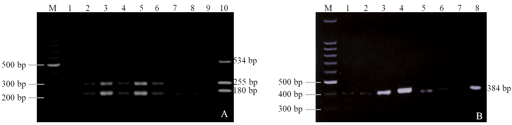
Figure 1. Agarose gels. (A) stx1, stx2 and hlyA genes, where M: molecular marker; lanes 1-8: E. coli isolated; -: negative control; +: positive control. (B) eaeA gene, where M: molecular marker; lanes 1-6 E. coli isolated; -: negative control; +: positive control
ERIC-PCR analysis
ERIC-PCR banding pattern analysis revealed 100% genetic diversity among the 13 E. coli isolates, with no identical genetic profiles observed. The isolates were differentiated into 13 unique patterns, clustering with an overall similarity index of 50%. Using a 50% similarity cut-off, four clusters were identified. Clusters 1, 2 and 3 each consisted of a single isolate (Mc9, McS2 and McS7, respectively), showing low similarity to the remaining isolates. Cluster 4 comprised 10 isolates (McS6, McS5, McS9, Mc8, Mc5, Mc10, Mc6, Mc3, Mc7 and Mc2), with internal similarity levels ranging from 18% to 54% (Figure 2).
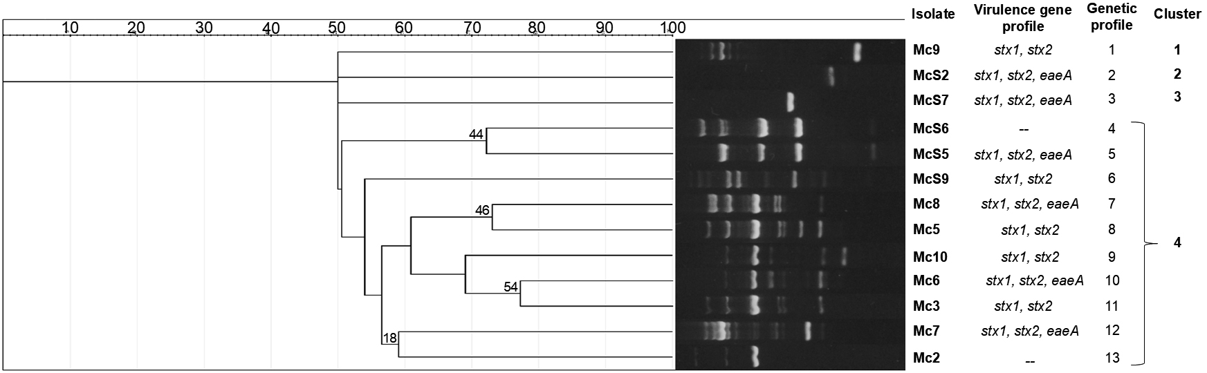
Figure 2. Dendrogram based on ERIC-PCR profiles of Escherichia coli isolated from ground beef in Hidalgo, Mexico
Beef production and consumption are essential to global food security. In this context, implementing practices that ensure meat safety is critical to reducing the occurrence of foodborne diseases and the mortality associated with them.33 Because of its high susceptibility to contamination during processing, ground beef represents an important vehicle for transmitting pathogenic agents. Thus, evaluating its microbiological load is key to understanding its role in foodborne disease incidence and the risks linked to contaminated meat consumption.34 This is particularly relevant given that meat can harbor pathogens such as STEC, an etiological agent of high prevalence and major public health concern. In the present study, an 80% prevalence of STEC was found in ground beef samples, a figure that aligns with previous reports from Mexico, where a 68% prevalence was documented in samples from Sonora.35 By contrast, lower prevalence rates have been reported elsewhere: 33.3% in Colombia,36 8.7% in Italy,37 and 9.72% in Vietnam.38 These differences may be attributed to variations in production practices, processing conditions, hygiene standards, the effectiveness of sanitary control systems, cold-chain infrastructure, or the detection methods employed. Indeed, the prevalence of STEC is known to vary across regions and depends strongly on methodological differences among studies. Nevertheless, the comparatively high prevalence reported here highlights a relevant public health risk and suggests possible deficiencies in manufacturing practices and sanitary conditions at butcher shops in the region studied. STEC contamination of meat is a multifactorial process that can occur at any stage of production. However, major contamination sources include cattle themselves, processing equipment, and workers involved at different points along the production chain.39,40 Ground beef is particularly susceptible to microbial contamination due to the grinding process, which facilitates cross-contamination through contact with equipment, mixing of cuts from different carcasses, and handling by workers exposed to multiple potentially contaminated surfaces.41-43 Of the 13 isolates analyzed in this study, 11 carried the stx1 and stx2 genes, confirming the pathogenic potential of these STEC strains. Shiga toxin, encoded by these genes, is the principal virulence factor of this pathotype and the causative agent of HUS. The epidemiological importance of this finding is considerable because Shiga toxin has been implicated in multiple serious outbreaks throughout history. For instance, the first reported outbreak of HUS caused by STEC occurred in 1982 in Oregon and Michigan, United States. Between that year and 2012, 653 cases of HUS and 73 STEC-associated deaths were documented in the United States.44 A major outbreak in Germany in 2011 resulted in 800 HUS cases, including 90 in children.45 In Argentina in 2019, 319 cases of HUS were reported, mainly affecting the child population.46 The virulence of STEC is further enhanced by other factors, such as intimin, encoded by the eaeA gene, which mediates adherence to enterocytes and is frequently investigated in diarrheal outbreaks. For example, eaeA has been detected in 18% of stool samples from children with diarrhea in Mexico City,47 in 10% of infant stool samples in Nigeria,48 and in 22.2% of stool samples from both children and adults with diarrhea in India.49 The detection of virulence genes is therefore essential for the identification and classification of E. coli. It facilitates both diagnosis and treatment, as well as predictions about strain pathogenicity and disease severity helping to distinguish between mild and severe infections.14 Monitoring STEC in foods such as meat is thus of critical importance because it represents a significant reservoir of this pathogen and is frequently implicated in epidemic outbreaks. The ERIC-PCR technique provided insight into the genetic diversity of the isolates through the analysis of their banding patterns. Our results revealed complete genetic diversity (100%) among the 13 isolates, with 13 unique patterns and no clones detected. These findings differ from those of other studies. For example, one investigation of 120 E. coli strains from animal feces identified only 10 genotypes, with no clones detected.50 Another study of 12 isolates from poultry feces reported 11 distinct genotypes, also without clonal patterns.51 By contrast, research on patients with urinary tract infections identified 12 clonal patterns among 20 isolates, reflecting greater genetic relatedness.52 The genetic variability of E. coli strains is well established and is driven by factors such as dispersal, recombination, mutation, and horizontal gene transfer.53-55 In this regard, ERIC-PCR is a valuable epidemiological tool, offering precise strain genotyping with high sensitivity and discriminatory power. It stands out for its reproducibility, low cost, and speed.51 Its use in subtyping enteric bacteria is essential for outbreak detection56 and for monitoring food contamination, as demonstrated in the present study.
This study confirmed the contamination of ground beef with STEC, evidenced by the detection of the virulence genes stx1, stx2, and eaeA in most of the isolates analyzed, indicating a high pathogenic potential. These findings position ground beef as a significant reservoir of pathogenic strains capable of transmission to humans through the consumption of contaminated products. ERIC-PCR banding pattern analysis revealed high genetic diversity and an absence of clonality among the isolates, suggesting multiple contamination sources. These results emphasize not only the need to improve hygienic and sanitary conditions at points of sale but also the importance of implementing continuous epidemiological surveillance strategies. Furthermore, promoting additional research to characterize associated risk factors will be essential for strengthening preventive measures and safeguarding public health in the region and in comparable contexts.
ACKNOWLEDGMENTS
None.
CONFLICT OF INTEREST
The authors declare that there is no conflict of interest.
AUTHORS’ CONTRIBUTION
VMMJ, VVS, NERR conceptualized the study and designed the experiments. APCL performed the experiments, analyzed the results and wrote the manuscript. FRGDA and JIOL supervised and edited the manuscript. APCL, VMMJ, VVS, NERR, RFGA and JIOL read and approved the final manuscript for publication.
FUNDING
None.
DATA AVAILABILITY
All datasets generated or analyzed during this study are included in the manuscript.
ETHICS STATEMENT
Not applicable.
- Secher T, Brehin C, Oswald E. Early settlers: which E. coli strains do you not want at birth? Am J Physiol Gastrointest Liver Physiol. 2016;311(1):G123-G129.
Crossref - Blount ZD. The unexhausted potential of E. coli. Elife. 2015;4(e05826):1-12.
Crossref - Petersen F, Hubbart JA. Physical factors impacting the survival and occurrence of Escherichia coli in secondary habitats. Water. 2020;12(6):1-15.
Crossref - Munekata PES, Pateiro M, Rodriguez-Lazaro D, Dominguez R, Zhong J, Lorenzo JM. The role of essential oils against pathogenic Escherichia coli in food products. Microorganisms. 2020;8(6):1-16.
Crossref - Jimenez MR, Gudino LFG, Loeza JAA, Lara PDL. Molecular characterization of antibiotic resistant Escherichia coli isolated from bovine mastitis in Michoacán, Mexico. Rev Mex Cienc Pecu. 2017;8(4):387-396.
Crossref - Kolenda R, Burdukiewicz M, Schierack P. A systematic review and meta-analysis of the epidemiology of pathogenic Escherichia coli of calves and the role of calves as reservoirs for human pathogenic E. coli. Front Cell Infect Microbiol. 2015;5:23.
Crossref - Riley LW. Distinguishing pathovars from nonpathovars: Escherichia coli. Microbiol Spectr. 2020;8(4):10.
Crossref - Gomes TAT, Elias W, Scaletsky ICA, et al. Diarrheagenic Escherichia coli. Braz J Microbiol. 2016;47(suppl 1):3-30.
Crossref - Newell DG, La Ragione RM. Enterohaemorrhagic and other Shiga toxin-producing Escherichia coli (STEC): Where are we now regarding diagnostics and control strategies? Transbound Emerg Dis. 2018;65(suppl 1)49-71.
Crossref - Majowicz SE, Scallan E, Jones-Bitton A, et al. Global incidence of human Shiga-toxin E. coli infections and deaths: a systematic review and knowledge synthesis. Foodborne Pathog Dis. 2014;11(6):447-455.
Crossref - FAO, WHO. Control measures for Shiga toxin-producing Escherichia coli (STEC) associated with meat and dairy products – Meeting report. Microbiol Risk Assess Ser No 39 Rome. 2022:1-220.
Crossref - Bryan A, Youngster I, McAdam A. Shiga Toxin Producing Escherichia coli. Clin Lab Med. 2015;35(2):247-272.
Crossref - Velez MV, Colello R, Etcheverria AI, Padola NL. Escherichia coli productora de toxina Shiga: el desafio de adherirse para sobrevivir. Rev Argent Microbiol. 2023;55(1):100-107.
Crossref - Pakbin B, Brück WM, Rossen JWA. Virulence factors of enteric pathogenic Escherichia coli: A review. Int J Mol Sci. 2021;22(18):9922.
Crossref - Gebisa ES, Gerasu MA, Leggese DT. A review on virulence factors of Escherichia coli. Anim Vet Sci. 2019;7(3):83-93.
Crossref - Moeinirad M, Douraghi M, Rahimi-Foroushani A, et al. Molecular characterization and prevalence of virulence factor genes of Shiga toxin-producing Escherichia coli (STEC) isolated from diarrheic children. Gene Reports. 2021;25:1-6.
Crossref - Onyeka LO, Adesiyun AA, Keddy KH, Madoroba E, Manqele A, Thompson PN. Shiga toxin-producing Escherichia coli contamination of raw beef and beef-based ready-to-eat products at retail outlets in Pretoria, South Africa. J Food Prot. 2020;83(3):476-484.
Crossref - Bantawa K, Rai K, Limbu DS, Khanal H. Food-borne bacterial pathogens in marketed raw meat of Dharan, eastern Nepal. BMC Res Notes. 2018;11(1):618.
Crossref - Abayneh M, Tesfaw G, Woldemichael K, Yohannis M, Abdissa A. Assessment of extended-spectrum β-lactamase (ESBLs) – producing Escherichia coli from minced meat of cattle and swab samples and hygienic status of meat retailer shops in Jimma town, Southwest Ethiopia. BMC Infect Dis. 2019;19(1):897.
Crossref - Manyi-Loh CE, Lues R. A South African perspective on the microbiological and chemical quality of meat: plausible public health implications. Microorganisms. 2023;11(10):284
Crossref - Cobos A, Diaz O. Chemical Composition of Meat and Meat Products. In: Cheung P, Mehta B (eds.). Handbook of Food Chemistry. Springer-Verlag, Berlin, Heidelberg 2015:471-510.
Crossref - Omer MK, Alvarez-Ordonez A, Prieto M, Skjerve E, Asehun T, Alvseike OA. A systematic review of bacterial foodborne outbreaks related to red meat and meat products. Foodborne Pathog Dis. 2018;15(10):598-611.
Crossref - Currie A, Honish L, Cutler J, et al. Outbreak of Escherichia coli O157:H7 infections linked to mechanically tenderized beef and the largest beef recall in Canada, 2012. J Food Prot. 2019;82(9):1532-1538.
Crossref - Warmate D, Onarinde BA. Food safety incidents in the red meat industry: A review of foodborne disease outbreaks linked to the consumption of red meat and its products, 1991 to 2021. Int J Food Microbiol. 2023;398:110240.
Crossref - Chung SM, Hellberg RS. Effects of poor sanitation procedures on cross-contamination of animal species in ground meat products. Food Control. 2020;109:1-6.
Crossref - Cordero-Lopez AP, Vega-Sanchez V, Martinez-Juarez VM, Olave-Leyva JI, Gomez-De Anda FR, Rodriguez NER. Antimicrobial resistance of Escherichia coli isolated from ground beef in Huasca de Ocampo, Hidalgo, Mexico. Trop Anim Sci J. 2025;48(1):75-82.
Crossref - Ribeiro Jr. JC, Tamanini R, Soares BF, et al. Efficiency of boiling and four other methods for genomic DNA extraction of deteriorating spore-forming bacteria from milk. Semina: Ciencias Agrarias. 2016;37(5):3069-3078.
Crossref - Paton AW, Paton JC. Detection and Characterization of Shiga toxigenic Escherichia coli by using Multiplex PCR Assays for stx1, stx2, eaeA, enterohemorrhagic E. coli hlyA, rfb O111, and rfb O157. J Clin Microbiol. 1998;36(2):598-602
Crossref - Okechukwu EC, Amuta EU, Gberikon GM, et al. Molecular identification of virulence genes of Escherichia coli isolated from cow milk and its products in Abuja, Nigeria. Microbiol Res J Int. 2020;30(6):11-18.
Crossref - Versalovic J, Koeuth T, Lupski J. Distribution of repetitive DNA sequences in eubacteria and application to fingerprinting of bacterial genomes. Nucleic Acids Res. 1991;19(24):6823-6831.
Crossref - Ardakani AM, Ranjbar R. Molecular typing of uropathogenic E. coli strains by the ERIC-PCR method. Electron Physician. 2016;8(4):2291-2296.
Crossref - Heras J, Dominguez C, Mata E, et al. GelJ – a tool for analyzing DNA fingerprint gel images. BMC Bioinformatics. 2015;16:270.
Crossref - Fung F, Wang HS, Menon S. Food safety in the 21st century. Biomed J. 2018;41(2):88-95.
Crossref - Ollinger M, Houser M. Ground beef recalls and subsequent food safety performance. Food Policy. 2020;97:1-15.
Crossref - Anduro-Jordan JA, Maldonado-Mendoza IE, Figueroa-Lopez AMT, et al. STEC non-0157 strains in meat from Southern Sonora, Mexico and their antibiotic resistance. Vet Mexico OA. 2022;9:1-16.
Crossref - Giraldo-Rubio V, Arango-Gil BS, Granobles-Velandia CV. First report of the prevalence of Shiga toxinproducing Escherichia coli in ground beef in Quindio, Colombia. Biomedica. 2023;43(4):474-482.
Crossref - Nobili G, Franconieri I, La Bella G, Basanisi MG, Salandra GL. Prevalence of Verocytotoxigenic Escherichia coli strains isolated from raw beef in southern Italy. Int J Food Microbiol. 2017;257:201-205.
Crossref - Duc HM, Ha CTT, Hoa TTK, Hung LV, Thang NV, Son HM. Prevalence, molecular characterization, and antimicrobial resistance profiles of Shiga toxin-producing Escherichia coli isolated from raw beef, pork, and chicken meat in Vietnam. Foods. 2024;13(13):1-11.
Crossref - Suryanto E, Syahlani SP, Airuni M. Good manufacturing practices implementation and microbiological quality of meat at the slaughterhouses in the Province of Bangka Belitung Islands. IOP Conf Ser Earth Environ Sci. 2019;387:1-6.
Crossref - Nekouei O, Checkley S, Waldner C, et al. Exposure to antimicrobial-resistant Escherichia coli through the consumption of ground beef in Western Canada. Int J Food Microbiol. 2018;272:41-48.
Crossref - Zhang Y, Schmidt JW, Arthur TM, Wheeler TL, Wang B. A comparative quantitative assessment of human exposure to various antimicrobial-resistant bacteria among U.S. Ground beef consumers. J Food Prot. 2021;84(5):736-759.
Crossref - Zerabruk K, Retta N, Muleta D, Tesfaye TA. Assessment of microbiological safety and quality of minced meat and meat contact surfaces in selected butcher shops of Addis Ababa, Ethiopia. J Food Qual. 2019;2019(1):1-9.
Crossref - Loukiadis E, Bieche-Terrier C, Malayrat C, Ferre F, Cartier P, Augustin JC. Distribution of Escherichia coli O157:H7 in ground beef: Assessing the clustering intensity for an industrial-scale grinder and a low and localized initial contamination. Int J Food Microbiol. 2017;250:75-81.
Crossref - Kim JS, Lee MS, Kim JH. Recent updates on outbreaks of shiga toxin-producing Escherichia coli and its potential reservoirs. Front Cell Infect Microbiol. 2020;10:273.
Crossref - Loos S, Aulbert W, Hoppe B, et al. Intermediate follow-up of pediatric patients with hemolytic uremic syndrome during the 2011 outbreak caused by E. coli O104:H4. Clin Infect Dis. 2017;64(12):1637-1643.
Crossref - Torti JF, Cuervo P, Nardello A, Pizarro M. Epidemiology and Characterization of Shiga Toxin-Producing Escherichia coli of Hemolytic Uremic Syndrome in Argentina. Cureus. 2021;13(8):1-6.
Crossref - Castro AM, Santos-Balbuena HF, Garcia-Garcia AE, Arzate-Barbosa. Association of diarrheagenic Escherichia coli with virotypes and sensitivity to antimicrobials in children of the Mexico City. Rev Med Hosp Gen Mex. 2019;82(2):87-97.
Crossref - David EE, Yameen MA, Igwenyi IO, et al. The frequency of virulent genes and antimicrobial resistance patterns of diarrheagenic Escherichia coli isolated from stools of children presenting with diarrhea in a tertiary hospital in Abakaliki, Nigeria. Int J One Heal. 2020;6(2):147-152.
Crossref - Natarajan M, Kumar D, Mandal J, Biswal N, Stephen S. A study of virulence and antimicrobial resistance pattern in diarrhoeagenic Escherichia coli isolated from diarrhoeal stool specimens from children and adults in a tertiary hospital, Puducherry, India. J Heal Popul Nutr. 2018;37(1):17.
Crossref - Ranjbar R, Tabatabaee A, Behzadi P, Kheiri R. Enterobacterial Repetitive Intergenic Consensus Polymerase Chain Reaction (ERIC-PCR) genotyping of Escherichia coli strains isolated from different animal stool specimens. Iran J Pathol. 2017;12(1):25-34.
Crossref - Sekhar MS, Sharif NM, Rao TS, Metta M. Genotyping of virulent Escherichia coli obtained from poultry and poultry farm workers using Enterobacterial Repetitive Intergenic Consensus-Polymerase Chain Reaction. Vet World. 2017;10(11):1292-1296.
Crossref - Lopez-Ramirez KL, Diaz-Maldonado KC, Vergara MA, et al. Patron de clonalidad mediante ERIC-PCR y REP-PCR de Escherichia coli y Klebsiella pneumoniae productores de betalactamasas de espectro extendido, aisladas de pacientes con infeccion urinaria intrahospitalaria. Hospital Regional Lambayeque, Peru. Horiz Med. 2018;18(2):11-18.
Crossref - Hanage WP. Not so simple after all: Bacteria, their population genetics, and recombination. Cold Spring Harb Perspect Biol. 2016;8(7):18069.
Crossref - Wiedenbeck J, Cohan FM. Origins of bacterial diversity through horizontal genetic transfer and adaptation to new ecological niches. FEMS Microbiol Rev. 2011;35(5):957-976.
Crossref - Bobay LM, Ochman H. Impact of Recombination on the Base Composition of Bacteria and Archaea. Mol Biol Evol. 2017;34(10):2627-2636.
Crossref - Bakhshi B, Afshari N, Fallah F. Enterobacterial Repetitive Intergenic Consensus (ERIC)-PCR analysis as a reliable evidence for suspected Shigella spp. Outbreaks. Brazilian J Microbiol. 2018;49(3):529-533.
Crossref
© The Author(s) 2025. Open Access. This article is distributed under the terms of the Creative Commons Attribution 4.0 International License which permits unrestricted use, sharing, distribution, and reproduction in any medium, provided you give appropriate credit to the original author(s) and the source, provide a link to the Creative Commons license, and indicate if changes were made.